AvicennaAspects

Triaging acute strokes: Emergency radiology gets AI support

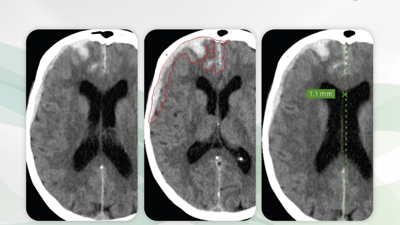
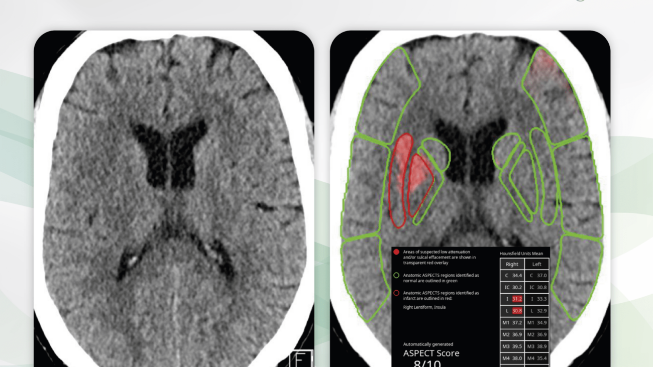
Non-enhanced cerebral CT of a 47-year-old patient with acute left sided paresthesia about 4 hours before the examination. Slightly low attenuation of the right lentiform nucleus and the right insular cortex is particularly visible when comparing the sides. No intracranial hemorrhage is visible (links). CINA-ASPECTS identifies the hypodensities and correctly assigns them to the lenticulostriatal territory (right, red). In addition, the results are tabulated, the ASPECTS is calculated as 8/10.

Time is Health: Our motto is particularly understandable when it comes to acute ischemic strokes. Every medical acute care unit knows what it means to accompany a large number of patients around the clock with the best diagnostic precision. Intracranial acute pathologies must be visualized quickly and safely.

It is with great pleasure that we present CINA-ASPECTS, an AI assistant for the detection of acute intracranial ischemia in CT studies.

Why CINA-ASPECTS matters abd how it works

CINA-ASPECTS was developed as a triage tool for emergency radiology. The AI assistant immediately reports suspected acute intracranial ischemia in CT studies and enables the prioritization of these patients. The AI assistant provides radiologists and emergency physicians with an outstanding presentation of the findings in words and images.

Any medical professional can use CINE-ASPECTS for each individual emergency patient by quickly and easily uploading cerebral CT studies to Radailogy. In medical institutions, this AI assistant can also do its work automatically in the background in order to fully exploit the desired triage potential.

Who benefits

The triage of acute ischemic strokes is essential for everyone involved, i.e., patients, clinicians and radiologists.

Our own experience at Radailogy

We were able to reproduce the statistical data of sensitivity of 76.6%, specificity of 88.7% and of accuracy of 87.0% given by the developer in our tests and partly worked out higher data for accuracy.

CINA-ASPECTS detects low attenuation of the supratentorial tissue and sulcal corticomedullar disorders. The eponymous topographical ASPECTS is used for the analysis: the supratentorial tissue is divided into ten regions of the middle cerebral artery supply per hemisphere to describe ischemia in one of the two hemispheres. The suspected pathological areas are marked in color and listed in a table with information on the mean density values ​​per region. From this, the ASPECTS for both hemispheres is calculated and given.

The developer cites as a limitation that older infarctions would not be correctly identified and describes the use of CINA-ASPECTS within 100 minutes of the clinical event as an essential inclusion criteria.

CINA-ASPECTS does not currently support the detection of posterior circulation ischemia (pc-ASPECTS). We expect this with the next upgrade.

We also evaluated CINA-ASPECTS together with two other AI assistants, CINA-ICH and CINA-LVO, which were developed to detect hemorrhagic and non-hemorrhagic strokes in CT studies. Find out more in our AI assistants!

The scientific evidence

Ayobi A, Chang P, Chow D, Filippi C, Quenet S, Tassy M, Chaibi Y. Validation of a Deep Learning AI-based Software for Automated ASPECTS Assessment. ECR 2023. DOI:           10.26044/ecr2023/C-19206

Data to upload to Radailogy

Non-enhanced cerebral CT studies of any CT scanner; axial reformations; minimal matrix size 256 x 256; maximal slice thickness 2.5 mm; tube current 100 kVp to 160 kVp (recommended 120 kVp to 140 kVp); soft tissue reconstruction kernel

AvicennaIvo

Cerebral artery occlusion: Emergency radiology gets AI support

CT angiography of the skull base arteries in a 68-year-old patient with worsening right sided paresthesia. In the coronal reformation, a contrast loss of the M2 segment of the left middle cerebral artery is be seen. Peripherally adjacent there is a large older ischemic defect (left). CINA-LVO correctly detects the artery occlusion (right, red box).

Time is Health: Our motto is particularly understandable when it comes to acute ischemic strokes. In about 80% they are caused by intracranial arterial thrombi. Rapid diagnosis using CT angiography is therefore essential.

It is with great pleasure that we present CINA-LVO, an AI assistant for the detection of intracranial artery occlusion in CT angiographies.

Why CINA-LVO matters and how it works

As a triage tool for emergency radiology, CINA-LVO reports suspected acute cerebral artery occlusions in CT angiographies and enables these patients to be prioritized. The AI assistant provides radiologists and emergency physicians with clear pictures.

Any medical professional can use CINA-LVO for each individual emergency patient by quickly and easily uploading CT angiographies of the skull base arteries to Radailogy. In medical institutions, this AI assistant can also do its work automatically in the background in order to fully benefit from its triage potential.

Who benefits

The triage of arterial ischemic strokes is essential for everyone involved, i.e., patients, clinicians and radiologists.

Our own experience at Radailogy

CINA-LVO (Large vessel occlusion) detects occlusions of the distal intracranial internal carotid artery and the M1 and M2 segments of the middle cerebral artery in CT angiographies of the skull base arteries. The suspected arteries are indicated in axial and coronal MIPs.

We were able to understand most of the statistical data given by the developer for sensitivity of 97.9%, specificity of 97.6% and accuracy of 97.7%. This correlates with the developer’s statement that occlusions less than 1.5mm are not recognized.

It is reasonable to use CINA-LVO together with CINA-ICH and CINA-ASPECTS, which were developed to detect hemorrhagic and non-hemorrhagic strokes in CT studies. Find out more in our AI assistants!

The scientific evidence

McLouth J, Elstrott S, Chaibi Y, Quenet S, Chang PD, Chow DS, Soun JE. Validation of a Deep Learning Tool in the Detection of Intracranial Hemorrhage and Large Vessel Occlusion. Front Neurol. 2021 Apr 29;12:656112.

Rava RA, Peterson BA, Seymour SE, Snyder KV, Mokin M, Waqas M, Hoi Y, Davies JM, Levy EI, Siddiqui AH, Ionita CN. Validation of an artificial intelligence driven large vessel occlusion detection algorithm for acute ischemic stroke patients. Neuroradiol J. 2021 Oct;34(5):408-417.

Schlossman J, Ro D, Salehi S, Chow D, Yu W, Chang PD, Soun JE. Headto head comparison of commercial artificial intelligence solutions for detection of large vessel occlusion at a comprehensive stroke center. Front Neurol. 2022 Oct 10;13:1026609.

Data to upload to Radailogy

CT angiographies of the skull base arteries of any CT scanner; axial reformations; minimal matrix size 512 x 512; maximal slice thickness 1.25 mm; tube current 80 kVp to 140 kVp (recommended 120 kVp to 140 kVp); 100 to 400 mAs; artery reconstruction kernel

AvicennaIPE

Acute pulmonary embolism: Emergency radiology gets AI support

CT angiography of the pulmonary arteries of a 58-year-old patient with acute respiratory distress and right-sided chest pain (left). There is a slight contrast sparing in the peripheral branching of the right lower lobe artery, affecting the lateral and dorsal segments. CINA-IPE correctly detects the embolization (right, red box).

Time is Health: Our motto is particularly understandable when it comes to acute pulmonary embolism. Diagnosis with CT angiography of the pulmonary arteries is therefore urgent. The automated early detection of positive findings helps every medical acute care unit that knows what it means to take care of a large number of patients around the clock.

It is with great pleasure that we present CINA-IPE, an AI assistant for the detection of acute pulmonary artery embolism in CT angiographies.

Why CINA-IPE matters and how it works 

As a triage tool for emergency radiology, CINA-IPE reports suspected thrombosis of the pulmonary arteries in CT angiographies and enables these patients to be prioritized. The AI assistant provides radiologists and emergency physicians with a clear visual presentation of findings.

Any medical professional can use CINA-IPE for each individual emergency patient by quickly and easily uploading CT angiographies of the pulmonary arteries to Radailogy. In medical institutions, this AI assistant can also do its work automatically in the background in order to fully benefit from its triage potential.

Who benefits

The triage of acute pulmonary embolism is essential for everyone involved, i.e., patients, clinicians and radiologists.

Our own experience at Radailogy

CINA-IPE detects emboli of the central and paracentral pulmonary arteries with excellent certainty. The suspected arteries are labeled in axial MIPs.

We were able to confirm the statistical data given by the developer of sensitivity of 86.6%, specificity of 92.7% and accuracy of 90.0% in our clinical tests. We recognized uncertainties in the detection of peripheral thrombosis. This correlates with the developer’s statement that subsegmental artery emboli are not detected.

The scientific evidence

Grenier PA, Ayobi A, Quenet S, Tassy M, Marx M, Chow DS, Weinberg BD, Chang PD, Chaibi Y. Deep Learning-Based Algorithm for Automatic Detection of Pulmonary Embolism in Chest CT Angiograms. Diagnostics. 2023;13(7), 1324

Schlossman J, Salehi S, Weinberg B, Chow D, Tassy M, Quenet S, Ayobi A, Chaibi Y, Chang P. Validation of a Deep Learning Tool for Automatic Pulmonary Embolism Detection. Am J Respir Crit Care Med. 2023;207:A2607

Data to upload to Radailogy

CT angiographies of the pulmonary arteries of any CT scanner; axial reformations; minimal matrix size 512 x 512; maximal slice thickness 2.5 mm; contrast enhancement of the central pulmonary arteries minimum 100 HU (recommended minimum 130 HU); soft tissue reconstruction kernel

AvicennaICH

Acute cerebral hemorrhage: Emergency Radiology gets AI support

Non-enhanced cerebral CT of a 46-year-old patient after high-speed trauma. There is an acute subdural hemorrhage along the right frontal and parietal hemisphere (left). In particular, it is frontally surrounded by incipient edema and a slight contralateral midline shift can be seen. There is no intraventricular hemorrhage. There is no evidence of a parafalcine herniation.

CINA-ICH correctly and fully detects intracranial hemorrhage (middle). Bleeding volume is reported within the CT series and also tabulated (not shown). The slight contralateral midline shift is precisely measured (right).

Time is Health: Our motto is particularly understandable when it comes to head injuries and strokes. Every medical acute care unit knows what it means to accompany a large number of patients around the clock with the best diagnostic precision. Intracranial acute pathologies must be visualized quickly and safely.

It is with great pleasure that we present CINA-ICH, an AI assistant for intracranial hemorrhage detection in CT studies.

Why CINA-ICH matters and how it works

CINA-ICH was developed as a triage tool for emergency radiology. The AI assistant promptly reports suspected intracranial hemorrhages in CT scans and enables prioritization of these patients. The AI assistant provides radiologists and emergency physicians with an outstanding presentation of the findings in words and images.

Any medical professional can use CINA-ICH for each individual emergency patient by quickly and easily uploading cerebral CT studies to Radailogy. In medical institutions, this AI assistant can also do its work automatically in the background in order to fully benefit from its triage potential.

Who benefits

The triage of traumatic brain injury and hemorrhagic stroke is essential for everyone involved, i.e., patients, clinicians and radiologists.

Our own experience at Radailogy

We were able to study the performance of CINA-ICH in detail in extensive test series. On the one hand, we were convinced by the quick and clear presentation of findings and, on the other hand, by the low false-negative and false-positive rates in the detection of intracranial bleeding. This is consistent with the statistical data provided by the developer of sensitivity and specificity of more than 90% each, measured on the total cohorts.

The results are reported in detail. Bleeding is plotted in axial CT volumes. In addition, the data are processed in tabular form and include the parenchymal, sub- and epidural, subarachnoid and intraventricular bleeding volumes. Furthermore, midline shifts are measured.

The manufacturer describes the smallest bleeding with a volume of less than 3 ml and non-acute haemorrhage as a possible false-negative detection. At Radailogy, we have not observed any missed bleeding in our clinical testing.

We also evaluated CINA-ICH together with two other AI assistants, CINA-ASPECTS and CINA-LVO, which were developed to detect non-hemorrhagic strokes in CT studies. Find out more in our AI assistants!

The scientific evidence

McLouth J, Elstrott S, Chaibi Y, Quenet S, Chang PD, Chow DS, Soun JE. Validation of a Deep Learning Tool in the Detection of Intracranial Hemorrhage and Large Vessel Occlusion. Front Neurol. 2021 Apr 29;12:656112.

Rava RA, Seymour SE, LaQue ME, Peterson BA, Snyder KV, Mokin M, Waqas M, Hoi Y, Davies JM, Levy EI, Siddiqui AH, Ionita CN. Assessment of an Artificial Intelligence Algorithm for Detection of Intracranial Hemorrhage. World Neurosurg. 2021 Jun;150:e2 09 e217.

Data to upload to Radailogy

Non-enhanced cerebral CT studies of any CT scanner; axial reformations; minimal matrix size 256 x 256; maximal slice thickness 5 mm; tube current 100 kVp to 160 kVp (recommended 120 kVp to 140 kVp); soft tissue reconstruction kernel

BoneviewMeasurements blog

Automated measurement of body axes on X-ray images

Frontal radiography of both forefeet of a 44-year-old patient. On the left (on the right in the picture) there is a clear hallux valgus deformity with an increase in the MTP joint angle I and the interdigital angle I/II. There is a mild secondary arthrosis of MTP joint I. There is moderate arthrosis in MTP joint II. Along the Lisfranc’s and the Chopart’s joints, there is a normal finding, apart from a small external tibial bone as a normal variant.

On the right (left in the picture) there is a normal finding of the forefoot, apparently subsequently to the osteomy of the first metatarsal bone.

BoneView Measurments enables the precise calculation of the joint angles and supports the radiological diagnosis with the angle measurements drawn in.

The measurement of body axes is one of the indispensable domains of radiology. At the same time, it places high demands on the precise description of the measured values ​​and thus on the available working time of radiologists.

It is with great pleasure that we present BoneView Measurements, an AI assistant for the automated measurement of body axes on X-ray images.

Why BoneView Measurments matters and how it works

BoneView Measurements is a fully automated AI assistant for measuring body axes. Its application minimizes the variability of the measurement results and ensures reproducibility. The AI assistant facilitates the interpretation of the findings thanks to its easy-to-read display.

Who benefits

Patients, clinicians and radiologists through the detailed measurement of body axes on X-ray images. BoneView Measurements can also be safely and easily integrated into the daily workflow.

Our own experience at Radailogy

The measurements work great for the spine, pelvis, hips, full leg scans and forefoot.

Any medical professional can request the measurement of body axes for each individual patient through the quick and easy upload to Radailogy. Our customers in telemedicine also use BoneView Measurements as a standard in daily practice to optimize their workflow.

The scientific evidence

Lassalle L, Regnard NE, Ventre J, Marty V, Clovis L, Zhang Z, Guermazi A, Laredo JD. Automated feet measurements using an artificial intelligence-based software. In press

Lassalle L, Regnard NE, Ventre J, Marty V, Clovis L, Zhang Z, Guermazi A, Laredo JD. Automated full-leg measurements using an artificial intelligence-based software. In press

Lassalle L, Regnard NE, Tran A, Ventre J, Marty V, Clovis L, Zhang Z, Guermazi A, Laredo JD. Automated hip measurements using an artificial intelligence-based software. In press

Data to upload to Radailogy

Digital radiography of the spine, pelvis, hips, full leg radiographs and forefoot from the age of three, depending on the anatomical region

bone age 1

AI for accurate bone age diagnosis

Radiography of the left hand a.p. of a male with the chronological age of nine years and three months. BoneView Bone Age calculates bone age using the Greulich & Pyle atlas method as seven years and ten months, standard deviation 10.74 months.

Bone age determination is one of the indispensable domains of radiology. At the same time, it places high demands on the clear description of the measured values.

It is with great pleasure that we present BoneView Bone Age, an AI assistant for pediatric bone age determination using hand radiographs.

Why BoneView Bone Age matters and how it works

BoneView Bone Age is an AI assitant for pediatric bone age determination according to the Greulich & Pyle atlas method for the age group from three to 17 years. The chronological age is compared with the AI ​​measurement data. In addition, the standard deviation is given in months.

Any medical professional anywhere in the world can request bone age analysis at any time by quickly and easily uploading hand radiographs to Radailogy. BoneView Bone Age offers the convenience and efficiency to simplify radiology workflows.

Who benefits

Patients, clinicians, and radiologists with accurate bone age diagnosis.

Our own experience at Radailogy

Any medical professional anywhere in the world can request bone age analysis at any time by quickly and easily uploading radiographs of the hand to Radailogy.

We found that BoneView Bone Age estimates are accurate and reasonably support our own radiological findings. We observed a very satisfactory inter- and intravariability with the integration of BoneView Bone Age.

The AI assistant is currently not suitable for determining bone age from the age of 18 and we are waiting for the AI ​​to evaluate the sternoclavicular joints with computed tomography.

The scientific evidence

Nguyen T, Pourchot A, Marty V, Ventre J, Regnard NE. Deep learning algorithm to predict Greulich and Pyle bone age. ESPR 2022 (June).

Data to upload to Radailogy

Digital radiography of the hand a.p. for the age group from three to 17 years.

Please upload only one radiography of the hand a.p. for your BoneView Bone Age order and do not combine BoneView Bone Age with another AI assistant within the same order!

BoneviewTrauma blog

Comprehensive AI fracture diagnosis of the peripheral skeleton

Radiographs of the left ankle frontal (left) and lateral (middle right) views of a 52-year-old patient with pain after a fall. BoneView Trauma correctly detects the nondisplaced longitudinal fracture of the distal fibula (middle left and right, yellow dashed boxes). In addition, the joint effusion is correctly reported in the lateral view (right, yellow box).

Radiographs of the peripheral skeleton are among the most common and important diagnostic methods for medical practices, medical institutes and hospitals. At the same time, they take up a large part of the available working time of radiologists, particularly because on average only about 10% of all radiographs show acute pathologies.

It is with great pleasure that we present BoneView Trauma, an AI assistant for fracture detection on X-ray images.

Why BoneView Trauma matters and how it works

BoneView Trauma detects traumatic lesions on X-rays. The AI diagnoses are available to all patients at any time of the day while providing the convenience and efficiency to streamline workflows. Fractures, effusions, dislocations, dislocations and malignant bone lesions are recognized. These pathologies are detected immediately after the X-rays are taken, even before the radiologist has seen the studies himself. These patients can be prioritized, diagnosed and treated accordingly.

Who benefits

Patients, clinicians and radiologists with a more detailed and accurate diagnosis and the reduced likelihood of missed therapy.

Our own experience at Radailogy

The high true positive rate of BoneView Trauma convinces us. Doubtful or borderline pathologies are also reported as such. In particular, the detection of joint effusions is currently unique. The clear delineation of pathologies in words and images supports the transfer of radiological knowledge to referring physicians and patients.

Any medical professional can request fracture analysis from this AI assistant for any individual patient by uploading it to Radailogy quickly and easily. Our telemedicine customers also use BoneView Trauma as a standard in their daily practice to streamline their workflow.

The scientific evidence

Cohen M, Puntonet J, Sanchez J, Kierszbaum E, Crema M, Soyer P, Dion E. Artificial intelligence vs. radiologist: accuracy of wrist fracture detection on radiographs. Eur Radiol. 2023 Jun;33(6):3974-3983

Duron L, Ducarouge A, Gillibert A, Lainé J, Allouche C, Cherel N, Zhang Z, Nitche N, Lacave E, Pourchot A, Felter A, Lassalle L, Regnard NE, Feydy A. Assessment of an AI Aid in Detection of Adult Appendicular Skeletal Fractures by Emergency Physicians and Radiologists: A Multicenter Cross-sectional Diagnostic Study. Radiology. 2021 Jul;300(1):120-129

Regnard NE, Lanseur B, Ventre J, Ducarouge A, Clovis L, Lassalle L, Lacave E, Grandjean A, Lambert A, Dallaudière B, Feydy A. Assessment of performances of a deep learning algorithm for the detection of limbs and pelvic fractures, dislocations, focal bone lesions, and elbow effusions on trauma X-rays. Eur J Radiol. 2022 Sep;154;110447

Data to upload to Radailogy

Digital radiography of the peripheral skeleton in two planes, for example a.p. and lateral or axial

ChestView blog

Rapid AI diagnosis of the chest X-ray

Chest radiograph of a 39-year-old patient (left) with symptoms of a lower respiratory tract infection. Small lung opacities are visible in projection to the left lower lung lobe. ChestView correctly identifies the pathology (yellow box). The result is also listed in a table (right). In addition to the fully automatic notification of the acute pathology, other threatening pathologies of the thorax are correctly evaluated as negative.

Chest radiography is used worldwide as one of the most common, if not the most common, radiological examinations in both acute care and elective medicine. Correct and reproducible reports and the communication of results from radiologists to patients and clinicians are of the utmost importance for everyone involved.

It is with great pleasure that we present ChestView, an AI assistant for chest radiography.

Why ChestView matters and how it works

ChestView is an expert-level AI assistant that has been extensively tested thanks to the joint work of our multidisciplinary team of developers and radiologist.

With ChestView essential pathologies of the thorax are diagnosed. The AI assistant was developed on the one hand to support triage in acute medicine and on the other hand to improve radiological work in terms of time savings and increased accuracy.

Who benefits

Patients, clinicians and radiologists by identifying the most important chest diseases with pictures and tables.

Our own experience at Radailogy

ChestView supports the detection of major thoracic pathologies. Although pneumonia is referred to as alveolar syndrome, the differential diagnosis has worked with a high degree of accuracy in our detailed tests. The presentation of the results by means of boxes and tabular description is helpful for radiological knowledge transfer to clinicians and patients.

The scientific evidence

Bennani S, Regnard NE, Lassalle L, Nguyen T, Malandrin C, Koulakian H, Khafagy P, Chassagnon G, Revel MP. Evaluation of radiologists’ performance compared to a deep learning algorithm for the detection of thoracic abnormalities on chest X-ray. In press

Data to upload to Radailogy

Digital radiographs of the chest for patients aged 15 and older