Rayscape blog 180223

Faster diagnoses for the chest X-ray in words and pictures

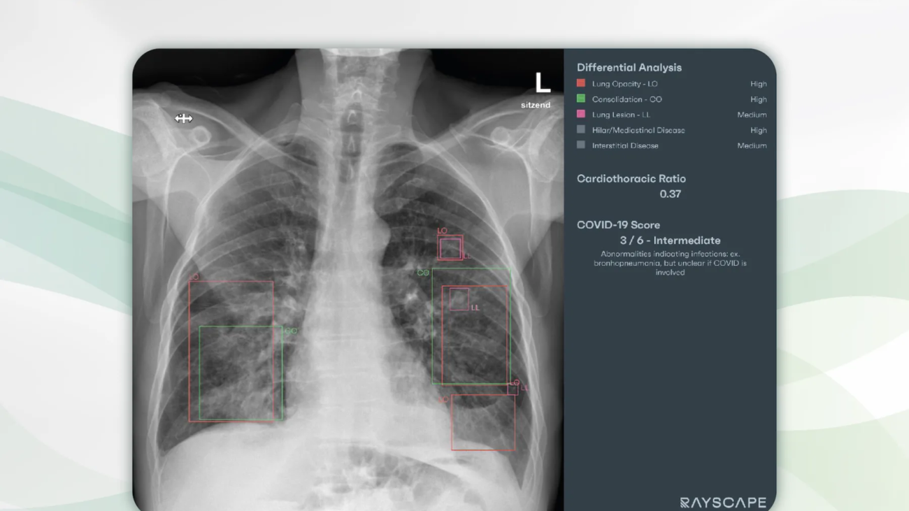
Chest radiography of a 65 years old patient with symptoms of lower respiratory tract infection. Multiple lung opacities and consolidations are visible in projection to the lower lung lobes as well as the middle lobe. Rayscape correctly identifies all consolidations (red) and opacities (green). Rayscape rates the probability for viral disease as intermediate (3 out of 6) and correctly suggests bronchopneumonia as the differential diagnosis. In addition, some small lung lesions are reported (pink) that were initially missed by the radiologists.

Chest radiography is one of the most common radiological examinations of all, and it is also an essential first-line imaging modality in hospitals and doctors´ offices. The precision in reporting and the communication of results from radiologists to patients and clinicians are the most important success factors for any optimal discussion of findings and therapy.

It is with great pleasure that we present Rayscape, an AI assistant for chest radiography.

What Rayscape is and how it works

Rayscape increases accuracy in detecting 17 major thoracic pathologies.

Thoracic findings are reported and visualized in tabular form. For each finding, the true positive rate is presented as a degree of probability versus differential diagnoses, especially for pulmonary nodules, pneumonia, and mediastinal pathologies. The clear presentation of findings is a welcome support for knowledge transfer from radiologists to patients and doctors.

Who benefits

Patients, clinicians and radiologists by identifying the most important chest diseases and injuries with a clear presentation of the findings in words and pictures.

Our own experience at Radailogy

We tested Rayscape for several years and brought it to life with the manufacturer. We reviewed the performance data and compared it to our own observations:

Rayscape supports the detection of pulmonary nodules, pulmonary consolidation, pulmonary edema, pulmonary emphysema, interstitial lung disease, tuberculosis, pneumothorax, pleural effusion, atelectasis, cardiomegaly, hilar and mediastinal pathologies, diaphragmatic abnormalities, fractures, scoliosis, catheters and drains. From our point of view, Rayscape achieves the best results in the detection of pneumonia, including viral pneumonia with probability grading from 1 to 6, pulmonary nodules, pneumothorax, pleural effusions, cardiac decompensation, consolidations and atelectasis.

The performance

Rayscape has a high level of accuracy in detecting 17 major chest pathologies. Area Under the Receiver Operating Characteristics (AUROC) is highest for tuberculosis (99.1), pneumothorax (97.4), pulmonary edema (94.7), consolidations (94.6), lowest for interstitial lung disease (81, 5), scoliosis (82), diaphragmatic anomalies (85.4), and hilar and mediastinal pathologies (87.7).

Data to upload to Radailogy

Digital radiography of the chest p.a. or a.p.


ColumboNeu web

MRI of the lumbar spine: CoLumbo with new features

T2 sagittal (left and middle) and T2 axial (right) MRI of the lumbar spine of a patient with back pain, lower limb weakness and paresthesia. Marked disc bulging (blue) and spinal canal stenosis at the level L2/L3, moderate disc bulging and spinal canal stenosis at the levels Th1-L2 and L4-S1. Left paramedian disc herniation (red), dural sac impingement (light blue) and recessal displacement of the left L4 nerve root at the level L3/L4. Foraminal stenosis and compression of the left foraminal nerves L3 and L4 (pink) at the levels L3-L5. Osteochondrosis Modic Type II at the levels L2/L3 and L4/L5.

With CoLumbo we have an excellent AI assistant for the assessment of the MRI of the lumbar spine. The latest release, now available on Radailogy, brings with it a variety of additional features for high-quality reporting of this common radiological examination. In particular, the description of foraminal stenoses and foraminal nerve impingement as well as the detection of osteochondrotic pathologies according to Modic are important.

It is with great pleasure that we present CoLumbo, an AI assistant for MRI of the lumbar spine.

Why CoLumbo is important and how it works

CoLumbo saves time and increases accuracy in the detection of the most common pathologies of the lumbar spine.

Lumbar spine MRI findings are reported and visualized. It assists in the knowledge transfer from radiologists to both patients and clinicians. In addition, all findings are written in comprehensive, standardized reports and can be used as an integral, automatically filling part of final documents.

Who benefits

Patients, clinicians, and radiologists by more detailed and more accurate diagnosis with subsequent decreased likelihood of suboptimal therapy or surgery. Accurate automatic measurements and clear colorful depiction reduce the need for measuring MRI findings by hand.

Our own experience at Radailogy

The AI ​​assistant supports the detection of disc herniation, disc bulging, central spinal canal stenosis, foraminal stenosis, nerve root impingement, reduced vertebral and disc height, hypo- and hyperlordosis, spondylolisthesis and pseudolisthesis as well as osteochondrotic pathologies according to Modic.

The scientific evidence

CoLumbo´s performance has been tested in clinical research with excellent results in accuracy for intervertebral disc detection and labeling (100%), for the detection of disc herniation (87%; 95% CI: 0.84, 0.89), extrusion (86%; 95% CI: 0.84, 0.89), bulging (76%; 95% CI: 0.73, 0.78), spinal canal stenosis (98%; 95% CI: 0.97, 0.99), nerve root compression (91%; 95% CI: 0.89, 0.92), and spondylolisthesis (87.61%; 95% CI: 85.26, 89.21), respectively.

Lehnen NC, Haase R, JFaber J, Rüber T, Vatter H, Radbruch A, Schmee FC. Detection of Degenerative Changes on MR Images of the Lumbar Spine with a Convolutional Neural Network: A Feasibility Study. Diagnostics 2021; 19;11(5):902

Data to upload to Radailogy

1.0-3.0 Tesla, T2 axial and sagittal 2D and 3D, slice thickness 3.45-5 mm

RayscapeLungCT

Oncological lung CT: Common, important and always time-consuming

Chest CT of a patient with a history of colorectal cancer. In the preliminary study dated June 13, 2023, two pulmonary nodules were visible in the left lower lobe (left). Rayscape Lung CT indicates the diameter and volume of each mass. In the follow-up CT dated September 29, 2023, these nodules progressed by 79.8% and 27.9%, respectively, in diameter and by 211.3% and 20.4%, respectively, in volume. In addition, three new nodules can be found in the lower lobe plane shown. Rayscape Lung CT marks these three lesions as new and indicates their diameter, volume and morphology (right).

Lung nodules occur in more than two million people per year in Europe alone. At the same time, the mortality rate from lung cancer worldwide is around two million annually. Their increase is expected to be around 30% in 10-year intervals. These few numbers alone illustrate the high level of responsibility that radiologists bear when interpreting computed tomography of the thorax. And the time-consuming nature of this task is particularly noticable when it comes to assessing lung nodules in each individual patient using their comparative studies.

It is with great pleasure that we present Rayscape Lung CT, an AI assistant for the detection of pulmonary nodules in CT studies.

Why Rayscape Lung CT matters and how it works

Rayscape Lung CT identifies pulmonary nodules from 3 to 30 mm in diameter. An important function is the automated comparison of nodules in the follow-up from CT study to CT study.

The results are reported in tabular form and visualized within the CT images. Each individual nodule is described with its exact location, diameter, volume and especially its morphology. These clear reports are an essential support for the transfer of knowledge from radiologists to patients and clinicians. Rayscape Lung CT can be used both for individual patients and in full-scale oncology departments.

Who benefits

Patients, clinicians and radiologists by the reliable detection of pulmonary nodules with clear reports in tables and images. In both screening and therapeutic oncology settings, the AI ​​assistant with its precise analysis enables timely and individual treatment.

Our own experience at Radailogy

Rayscape Lung CT has high values ​​for PPV, NPV and sensitivity of more than 97% each in our own patient population. At Radailogy, sensitivity in the correct detection of pulmonary nodules is over 93%. Nodules are precisely located and described using the parameters diameter, volume and morphology. If requested, up to three CT studies are compared in the follow-up setting. New nodules are reported as such. In particular, the automated comparison in follow-up lung CTs saves us an amazing amount of time for each individual patient every day and provides us with the desired additional security in the completeness of our findings.

Rayscape Lung CT performs a risk analysis for each study based on the established Fleischer criteria. We recognize this addition as useful, but we have not often used it in daily practice when discussing findings with clinicians and patients.

As described, the AI ​​assistant detects lung nodules with high reliability. We found that primary tumors larger than 30 mm in diameter are not described in detail in the oncological follow-up. However, this is not the goal of the software formulated by the manufacturer.

The scientific evidence

Tenescu A, Bercean BA, Avramescu C, Marcu M. Averaging Model Weights Boosts Automated Lung Nodule Detection on Computed Tomography. 13th International Conference on Bioscience, Biochemistry and Bioinformatics. 2023 ISBN 978-1-4503-9819-0.

Benta MM, Rasadean C, Ardelean PG, Barbulescu I, Birhala A, Bercean B, Avramescu C, Tenescu A, Birsasteanu F. Artificial intelligence in computed tomography – lung nodule analysis algorithm. ECR 2022 DOI 10.26044/ecr2022/C-17388.

Data to upload to Radailogy

Native or contrast enhanced CT studies of the chest of any CT scanner age and vendor; axial reformations; maximal slice thickness 3 mm; lung reconstruction kernel; patient age at least 16 years