AiraMed copy

Early detection of neurodegenerative diseases for a successful treatment

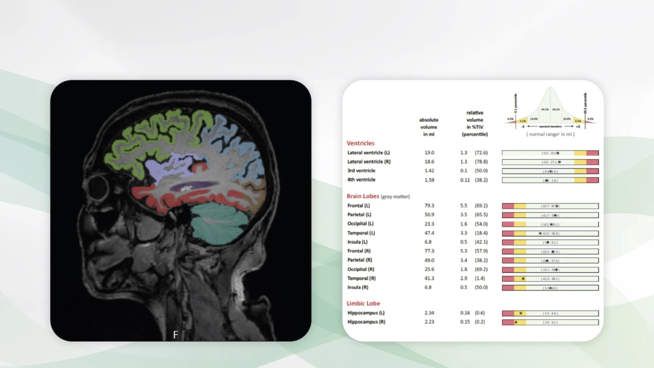
3D T1-weighted gradient-echo MRI of a 68 years old patient with mild symptoms of cognitive impairment. Routine MRI (not shown) revealed mild microangiopathic neurodegeneration. Brain volumetry was performed (left). AIRAscore identified a volume reduction of the right temporal lobe and bilateral hippocampal that exceeds age (right), suggestive for Alzheimer´s disease. Diagnosis was confirmed by cerebrospinal fluid analysis showing pathological beta amyloid ratio and elevated tau and phosphor-tau proteins.

For many people it remains unrecognized that even minor symptoms can indicate the development of dementia. They thus miss the important early diagnosis and the timely start of their individualized therapy and thus the best possible prognosis for many years. AI, together with brain MRI, can contribute to the earliest possible detection and treatment of neurodegenerative diseases.

It is with great pleasure that we present AIRAscore, an AI assistant for the precise diagnosis of neurodegenerative diseases.

What AIRAscore is and how it works

AIRAscore measures relevant biomarkers for the diagnosis and differential diagnosis of neurodegenerative diseases from MR images of the brain.

Global and regional brain volumes are accurately measured. The results are always compared with age- and gender-specific reference values. The measurements are many times more accurate than even specialists have been able to carry out in the past. Results that deviate from normal are clearly evident. The findings are reported in tabular form in bar graphs and support the knowledge transfer from radiologists to patients and physicians.

Who benefits

Patients, clinicians and radiologists through the early detection and differential diagnosis of neurodegenerative diseases. AIRAscore is also ideal for monitoring the progress of diagnoses made.

Our own experience at Radailogy

We have tested AIRAscore intensively and worked out many technological criteria with the manufacturer for optimal use with Radailogy.

We found a high correlation in the detection of macro- and microangiopathic changes and brain atrophy between our specialists and AIRAscore. In addition, AIRAscore was superior to the human observer in the precision of subtle regional findings in almost all tests. The AI assistant provides accurate volumes of all relevant anatomical structures of the cerebrum, cerebellum and brainstem. Early diagnosis was possible, especially for Alzheimer’s patients, and the correlation with clinical and laboratory data was also very high in the follow-up. AIRAscore can also be used for the differential diagnosis of multiple sclerosis and Parkinson’s disease.

The scientific environment

AIRAscore was developed in neuroscientific medical research. From our point of view, the AI assistant represents a bridge between innovative university research and direct applicability in everyday clinical practice.

Data to upload to Radailogy

1.5-3.0 Tesla MRI, native 3D T1-weighted gradient-echo sequences, slice thickness 1 mm, echo time ≤ 5 ms, flip angle ≤ 15°

PixelShine2_1

Computed tomography: The most important radiological method with improved image quality and reduced radiation dose

Low-dose chest CT of a patient with a pneumonic infiltrate of the middle lobe. Original image with 80kVp, 15 mAs, radiation dose 0.2 mGy (bottom left). Processing with Pixelshine (top left). A comparable image would be created with about 120 kVp, 150 mAs and a radiation dose of about 8 mGy. The radiation dose is reduced by more than 95% with PixelShine.

Low-dose CT of the abdomen. Original image with 120kVp, slice thickness 1.25 mm, radiation dose 1.7 mGy (bottom middle). Processing with Pixelshine (top middle). A comparable image would be created with a radiation dose of about 10 mGy. The radiation dose is reduced by more than 80% with PixelShine.

CT of the brain. Original image with 120kVp, slice thickness 0.625 mm, radiation dose 11 mGy (bottom right). Processing with Pixelshine (top right). A comparable image would be created with a radiation dose of about 40 mGy. The radiation dose is reduced by around 75% with PixelShine.

For several years, medicine has been generating significantly more radiation doses than the natural radiation from the cosmos and the earth ever did. The main reason for this is the constantly increasing radiological use of computed tomography (CT). Precisely because CT is and will remain essential for adequate patient care in almost all diagnostic areas, it is up to us to keep the long-documented radiation-induced cancer risk at the lowest possible level.

It is with great pleasure that we present PixelShine, an AI assistant for CT radiation dose reduction.

Why PixelShine matters

Both in the hospital and in the radiological institute, care is taken to ensure that each patient is only given the necessary radiation dose. However, these low-dose CT protocols almost always produce noisy images, and the CT studies are often difficult to interpret even for medical specialists. In addition, radiologists often have to read CT studies from CT machines from different vendors, which contributes to inconvenience and delays in the workflow.

PixelShine allows two things: Firstly, low-dose CT studies can be carried out for all patients in terms of optimal radiation protection, and PixelShine subsequently generates significantly improved quality from these noisy images, for example in obese patients. Secondly, the lifespan of CT scanners is extended by reducing the load on the CT tubes.

When and how to use PixelShine

PixelShine can be used for studies of any CT device age and vendor. This AI ​​assistant improves radiological precision by homogenizing the workflow.

PixelShine enables radiologists to read noisy CT studies with a high noise level of image noise in the best possible way, and the radiological quality meets the requirements for diagnostic validity.

Furthermore, hospitals and radiological institutes can carry out low-dose CT studies as standard, integrate PixelShine in post-processing and thus achieve consistently high image quality.

Who benefits

Patients, clinicians, radiologists and the management of hospitals and radiological institutes: care for all patients by minimizing the radiation dose, clear CT images, optimal assessment and discussion of findings, money savings by extending the lifespan of CT scanners.

Our own experience at Radailogy

Our customers send us CT studies to improve image quality with PixelShine and enable optimal diagnostic results. Both in individual cases through the simple upload to Radailogy, as well as as a standard in daily cooperation with our telemedicine.

Selection of scientific publications

Hata A, Yanagawa M, Yoshida Y, et al. Combination of Deep Learning–Based Denoising and Iterative Reconstruction for Ultra-Low-Dose CT of the Chest: Image Quality and Lung-RADS Evaluation. American Journal of Roentgenology. 2020;215(6):1321-1328.

Steuwe A, Weber M, Bethge OT, et al. Influence of a novel deep-learning based reconstruction software on the objective and subjective image quality in low-dose abdominal computed tomography. BJR. 2021;94(1117):20200677.

Brendlin AS, Plajer D, Chaika M, et al. AI Denoising Significantly Improves Image Quality in Whole-Body Low-Dose Computed Tomography Staging. Diagnostics. 2022;12(1):225.

Hasegawa A, Ishihara T, Thomas MA, Pan T. Noise reduction profile: A new method for evaluation of noise reduction techniques in CT. Medical Physics. 2022;49(1):186-200.

Nagaraj Y, de Jonge G, Andreychenko A, et al. Facilitating standardized COVID-19 suspicion prediction based on computed tomography radiomics in a multi-demographic setting. Eur Radiol. 2022;32(9):6384-6396.

Hasegawa A, Ishihara T, Thomas MA, Pan T. Noise reduction profile: A new method for evaluation of noise reduction techniques in CT. Medical Physics. 2022;49(1):186-200.

Data to upload to Radailogy

CT studies of any CT scanner age and vendor