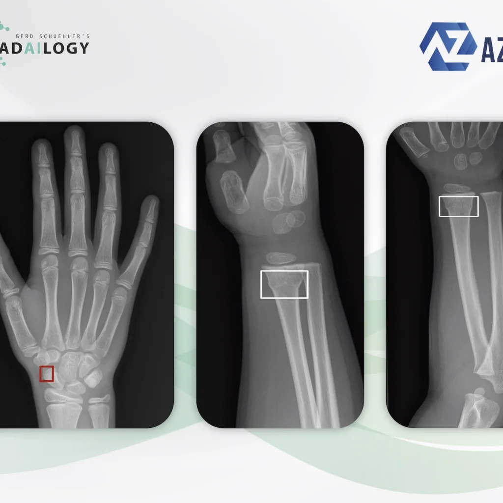
Radiography of the right hand a.p. of a 13-year-old patient with pain in the snuff box after a fall (left). Rayvolve correctly identifies the non-displaced fracture of the distal pole of the scaphoid. Copyright AZMed
Radiography of the right forearm and wrist a.p. (right) and oblique (middle) of a 4-year-old patient after a fall. Rayvolve correctly recognizes the torus fracture of the distal radius, which is rather rare for this patient age. Copyright AZMed
Traumatology plays an important role day and night in medical practices, medical institutes and hospitals. At the same time, it takes up a large proportion of the available working time for medical professionals. On average, only about 10% of all X-rays actually show fractures.
It is with great pleasure that we present Rayvolve, an AI assistant for fracture detection on X-ray images.
Why Rayvolve matters and how it works
Rayvolve supports rapid and reliable fracture diagnosis on X-ray images of the axial and peripheral skeleton.
Fracture findings are visualized in clear images. The manufacturer speaks of a time saving for radiologists of almost 30% and a significant improvement in diagnostic accuracy with a reduction in false negative results of more than 60%.
Any medical professional can use Rayvolve for each and every trauma patient. In medical institutions, this AI assistant can also do its work automatically in the background. As a result, fractures are detected immediately after the X-rays are taken, even before the radiologist has seen the studies themselves. These patients can be prioritized, diagnosed and treated accordingly.
Who benefits
Patients, clinicians and radiologists with a more detailed and accurate diagnosis and the reduced likelihood of missed therapy.
Our own experience at Radailogy
Any medical professional can request fracture analysis from this AI assistant for any individual patient by uploading it to Radailogy quickly and easily. Our telemedicine customers also use Rayvolve as a standard in their daily practice to streamline their workflow.
Our own results at Radailogy with a cohort of a few hundred patients are: sensitivity 93.5%, specificity 87.8%, positive predictive value 92%, negative predictive value 93%.
The scientific evidence
Dupuis M, Delbos L, Veil R, Adamsbaum C. External validation of a commercially available deep learning algorithm for fracture detection in children. Diagn Interv Imaging. 2022 Mar;103(3):151-159.
Date to upload to Radailogy
Digital radiography of a body region in two planes, for example a.p. and lateral or axial

