
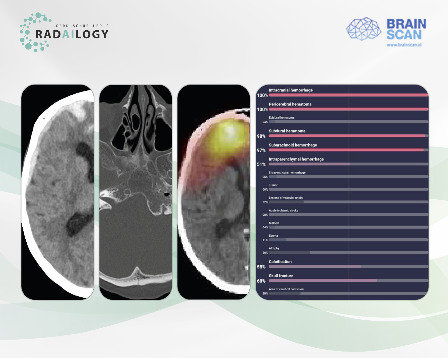
Non-enhanced cerebral CT of a 61-year-old patient after a motor vehicle accident. There is acute intraparenchymal, subdural, and subarachnoid hemorrhage along the right hemisphere (left) and a nondisplaced occipital skull fracture (middle left).
Brainscan CT detects and reports all details of intracranial hemorrhage and skull fracture with high accuracy as a heat map (middle right) and also tabulated (right).
Not every patient with symptoms of acute intracranial pathology has a corresponding CT correlate, but rather a chronic cerebral process – and vice versa. Therefore, the comprehensive detection of all intracranial pathologies is one of the main tasks of neurologists and radiologists, be it in acute medicine or in routine diagnostics.
It is with great pleasure that we present Brainscan CT, an AI assistant for the detection of acute and chronic intracranial pathologies in CT studies.
Why Brainscan CT matters and how it works
The AI assistant reports acute and chronic intracranial processes in CT scans. Using tables and heat maps, it gives neurologists, radiologists and emergency physicians a direct view of crucial brain pathologies.
Any medical professional can use Brainscan CT for each individual emergency patient by quickly and easily uploading cerebral CT studies to Radailogy. In medical institutions, this AI software can also do its work automatically in the background in order to fully benefit from its triage potential.
Any medical professional can use Brainscan CT by quickly and easily uploading cerebral CT studies to Radailogy. In acute medicine, this AI assistant can also be integrated into the workflow as a standard to fully utilize triage protocols.
Who benefits
Patients, clinicians and radiologists through the comprehensive detection of intracranial pathologies and the outstanding presentation of the results in tables and heat maps.
Our own experience at Radailogy
Brainscan CT detected intraparenchymal, subdural, epidural and subarachnoid hemorrhages with high accuracy in our test series. This corresponds to the performance data given by the manufacturer: sensitivity of an average of 85% with a specificity of at least 95%. Skull fractures were recognized well, even if the sensitivity is only 51% according to the manufacturer. Acute infarctions were detected sufficiently well, although the differentiation from chronic processes was not without exceptions, correlating with a sensitivity of almost 60% and a specificity of 99%. Other pathologies detected by Brainscan CT include: vascular lesions in general, leukoencephalopathy, edema, atrophy, calcifications, and tumors. We were not able to test the latter in detail.
The results are presented using tables. The presence of intracranial pathologies is given as a percentage – as an expression of probability – and is also represented with a bar chart. What initially takes some getting used to is that the specification of 51% already means the detection of individual lesions. The AI assistant produced clear results in the vast majority of our test series. We only observed statements of 50% probability in exceptions in which even radiologists would not naturally come to a clear opinion.
In the current version, Brainscan CT also shows intracranial hemorrhages and leukoencephalopathy using impressive heat maps. The results are delivered directly to the PACS as DICOM images.
Data to upload to Radailogy
Non-enhanced cerebral CT studies of any CT scanner for patients aged 18 and older; axial reformations; maximum matrix size 512 x 512; maximal slice thickness 5 mm; soft tissue reconstruction kernel