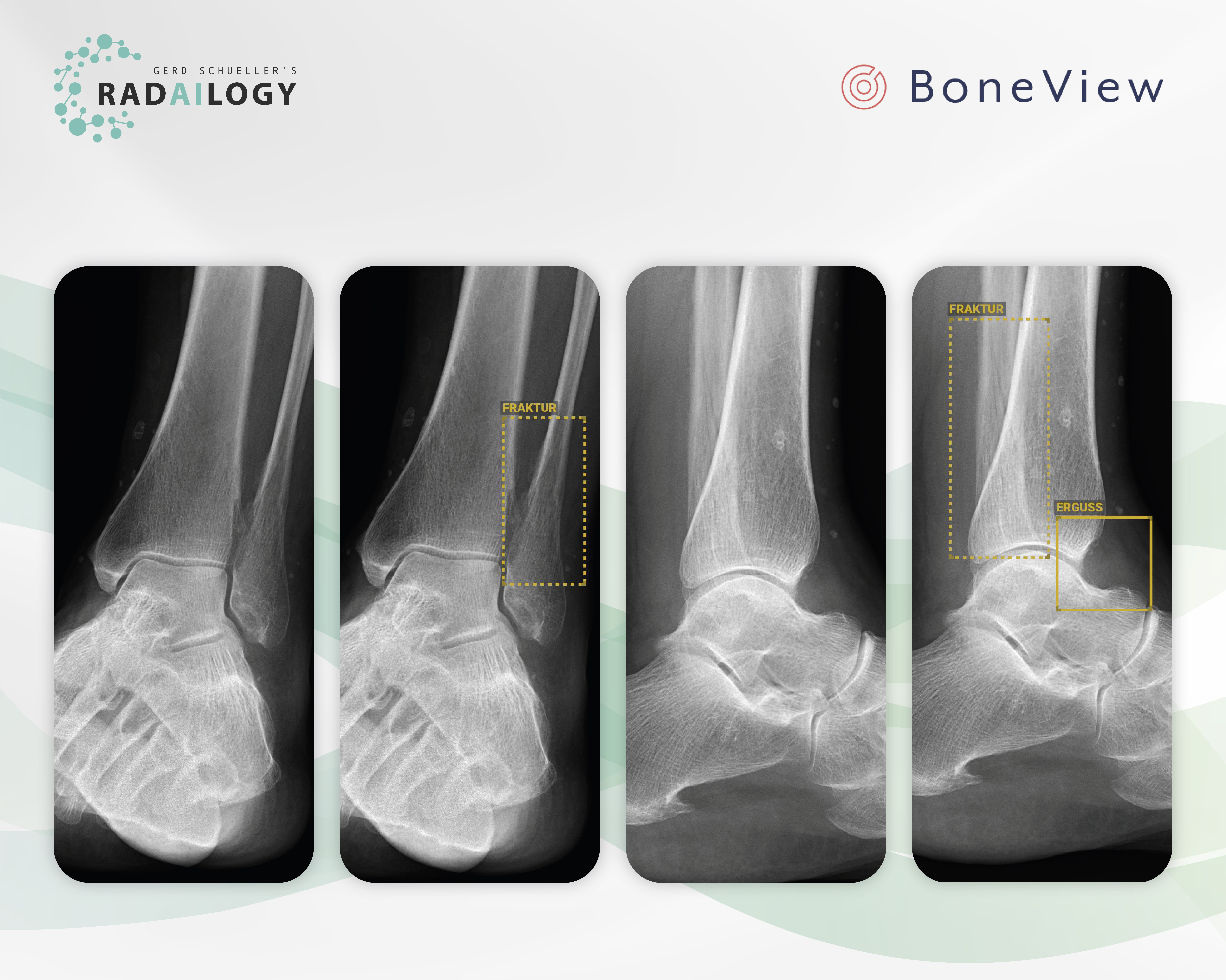
Radiographs of the left ankle frontal (left) and lateral (middle right) views of a 52-year-old patient with pain after a fall. BoneView Trauma correctly detects the nondisplaced longitudinal fracture of the distal fibula (middle left and right, yellow dashed boxes). In addition, the joint effusion is correctly reported in the lateral view (right, yellow box).
Radiographs of the peripheral skeleton are among the most common and important diagnostic methods for medical practices, medical institutes and hospitals. At the same time, they take up a large part of the available working time of radiologists, particularly because on average only about 10% of all radiographs show acute pathologies.
It is with great pleasure that we present BoneView Trauma, an AI assistant for fracture detection on X-ray images.
Why BoneView Trauma matters and how it works
BoneView Trauma detects traumatic lesions on X-rays. The AI diagnoses are available to all patients at any time of the day while providing the convenience and efficiency to streamline workflows. Fractures, effusions, dislocations, dislocations and malignant bone lesions are recognized. These pathologies are detected immediately after the X-rays are taken, even before the radiologist has seen the studies himself. These patients can be prioritized, diagnosed and treated accordingly.
Who benefits
Patients, clinicians and radiologists with a more detailed and accurate diagnosis and the reduced likelihood of missed therapy.
Our own experience at Radailogy
The high true positive rate of BoneView Trauma convinces us. Doubtful or borderline pathologies are also reported as such. In particular, the detection of joint effusions is currently unique. The clear delineation of pathologies in words and images supports the transfer of radiological knowledge to referring physicians and patients.
Any medical professional can request fracture analysis from this AI assistant for any individual patient by uploading it to Radailogy quickly and easily. Our telemedicine customers also use BoneView Trauma as a standard in their daily practice to streamline their workflow.
The scientific evidence
Cohen M, Puntonet J, Sanchez J, Kierszbaum E, Crema M, Soyer P, Dion E. Artificial intelligence vs. radiologist: accuracy of wrist fracture detection on radiographs. Eur Radiol. 2023 Jun;33(6):3974-3983
Duron L, Ducarouge A, Gillibert A, Lainé J, Allouche C, Cherel N, Zhang Z, Nitche N, Lacave E, Pourchot A, Felter A, Lassalle L, Regnard NE, Feydy A. Assessment of an AI Aid in Detection of Adult Appendicular Skeletal Fractures by Emergency Physicians and Radiologists: A Multicenter Cross-sectional Diagnostic Study. Radiology. 2021 Jul;300(1):120-129
Regnard NE, Lanseur B, Ventre J, Ducarouge A, Clovis L, Lassalle L, Lacave E, Grandjean A, Lambert A, Dallaudière B, Feydy A. Assessment of performances of a deep learning algorithm for the detection of limbs and pelvic fractures, dislocations, focal bone lesions, and elbow effusions on trauma X-rays. Eur J Radiol. 2022 Sep;154;110447
Data to upload to Radailogy
Digital radiography of the peripheral skeleton in two planes, for example a.p. and lateral or axial

