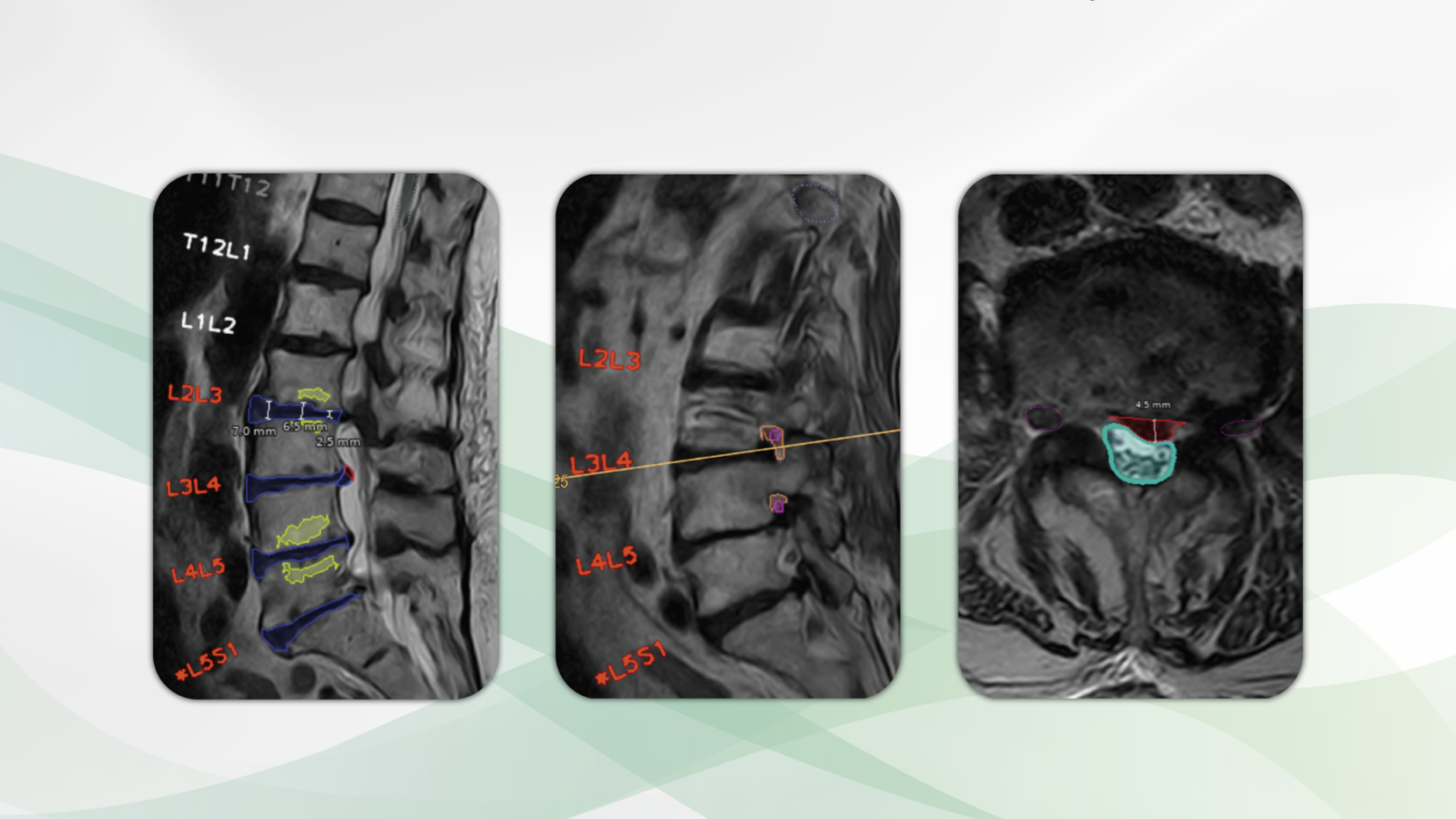
Rayscape blog 180223

Faster diagnoses for the chest X-ray in words and pictures

Chest radiography of a 65 years old patient with symptoms of lower respiratory tract infection. Multiple lung opacities and consolidations are visible in projection to the lower lung lobes as well as the middle lobe. Rayscape correctly identifies all consolidations (red) and opacities (green). Rayscape rates the probability for viral disease as intermediate (3 out of 6) and correctly suggests bronchopneumonia as the differential diagnosis. In addition, some small lung lesions are reported (pink) that were initially missed by the radiologists.

Chest radiography is one of the most common radiological examinations of all, and it is also an essential first-line imaging modality in hospitals and doctors´ offices. The precision in reporting and the communication of results from radiologists to patients and clinicians are the most important success factors for any optimal discussion of findings and therapy.

It is with great pleasure that we present Rayscape, an AI assistant for chest radiography.

What Rayscape is and how it works

Rayscape increases accuracy in detecting 17 major thoracic pathologies.

Thoracic findings are reported and visualized in tabular form. For each finding, the true positive rate is presented as a degree of probability versus differential diagnoses, especially for pulmonary nodules, pneumonia, and mediastinal pathologies. The clear presentation of findings is a welcome support for knowledge transfer from radiologists to patients and doctors.

Who benefits

Patients, clinicians and radiologists by identifying the most important chest diseases and injuries with a clear presentation of the findings in words and pictures.

Our own experience at Radailogy

We tested Rayscape for several years and brought it to life with the manufacturer. We reviewed the performance data and compared it to our own observations:

Rayscape supports the detection of pulmonary nodules, pulmonary consolidation, pulmonary edema, pulmonary emphysema, interstitial lung disease, tuberculosis, pneumothorax, pleural effusion, atelectasis, cardiomegaly, hilar and mediastinal pathologies, diaphragmatic abnormalities, fractures, scoliosis, catheters and drains. From our point of view, Rayscape achieves the best results in the detection of pneumonia, including viral pneumonia with probability grading from 1 to 6, pulmonary nodules, pneumothorax, pleural effusions, cardiac decompensation, consolidations and atelectasis.

The performance

Rayscape has a high level of accuracy in detecting 17 major chest pathologies. Area Under the Receiver Operating Characteristics (AUROC) is highest for tuberculosis (99.1), pneumothorax (97.4), pulmonary edema (94.7), consolidations (94.6), lowest for interstitial lung disease (81, 5), scoliosis (82), diaphragmatic anomalies (85.4), and hilar and mediastinal pathologies (87.7).

Data to upload to Radailogy

Digital radiography of the chest p.a. or a.p.


Featured-Image-Blog-2

AI brings patients and doctors together

Physicians depend on exam data from a variety of medical specialties to inform patients about their current situation and enable the best to heal them. 

Radiology and its fast and accurate diagnostics often present itself as the basis on which every successful therapy relies upon. The most important success factors are the precision in the reporting and the transmission of the results to patients and clinicians.

Patients and clinicians rightly want precise analyses presented in words and pictures. X-rays, CT and MR images are ideal for discussing findings. Here, clinicians depend on the results being clearly visible in order to show them to their patients without loss of information or quality.

There has been a 78% increase in radiological examinations since 2008, and the trend is rising. At the same time, healthcare systems are facing a significant shortage of medical specialists. That essential, precise transfer of knowledge from radiologists to patients and clinicians is often subject to daily time pressure.

Radailogy’s AI creates a better future. AI manufacturers know what requirements and expectations we have of their products. We test and perfect AI assistants hand in hand with its developers. At Radailogy, we care about everyone involved: patients, clinicians and radiologists. With every AI assistant we offer, we help save time, increase medical precision, and see and understand exam results clearly.

Columbo

Optimal lumbar spine MRI visualisation

T2 axial (upper left) and T2 sagittal (lower left) MRI of the lumbar spine of a patient with back pain, lower limb weakness and paresthesia. Left foraminal disc extrusion (red) at the level L4/L5 with impingement of the left L4 nerve (pink) and discrete recessal displacement of the left L5 nerve root (pink). T2 axial (upper right) and T2 sagittal (lower right) MRI of a patient with left lower limb weakness and severe back pain. Left paramedian disc herniation (red), dural sac compression (light blue), recessal displacement of the left L5 nerve root (pink). No foraminal nerve compression (pink). Grade I retrolisthesis.

The MRI of the lumbar spine is one of the most common radiological examinations. Four out of five people will have it at some point in their lives. The most important success factors for any optimal therapy are the precision in reporting and the transmission of the results from radiologists to patients and clinicians.

It is with great pleasure that we present CoLumbo, an AI assistant for MRI of the lumbar spine.

What CoLumbo is and how it works

CoLumbo saves time and increases accuracy in the detection of the most common pathologies of the lumbar spine.

Lumbar spine MRI findings are reported and visualized. It assists in the knowledge transfer from radiologists to both patients and clinicians. In addition, all findings are written in comprehensive, standardized reports and can be used as an integral, automatically filling part of final documents.

Who benefits

Patients, clinicians, and radiologists by more detailed and more accurate diagnosis with subsequent decreased likelihood of suboptimal therapy or surgery. Accurate automatic measurements and clear colorful depiction reduce the need for measuring MRI findings by hand.

Our own experience at Radailogy

We have scrunitely studied CoLumbo over many months. Here we share some of our results with you:

This AI assistant supports the detection of disc herniation, disc bulging, central spinal stenosis, nerve root impingement, reduced vertebral and disk height, hypo- and hyperlordosis as well as spondylolisthesis and pseudolisthesis. With the actual CoLumbo version, we recommend the specialist´s review of foraminal nerve root impingement.

The scientific evidence

CoLumbo´s performance has been tested in clinical research with excellent results in accuracy for intervertebral disc detection and labeling (100%), for the detection of disc herniation (87%; 95% CI: 0.84, 0.89), extrusion (86%; 95% CI: 0.84, 0.89), bulging (76%; 95% CI: 0.73, 0.78), spinal canal stenosis (98%; 95% CI: 0.97, 0.99), nerve root compression (91%; 95% CI: 0.89, 0.92), and spondylolisthesis (87.61%; 95% CI: 85.26, 89.21), respectively.

Lehnen NC, Haase R, JFaber J, Rüber T, Vatter H, Radbruch A, Schmee FC. Detection of Degenerative Changes on MR Images of the Lumbar Spine with a Convolutional Neural Network: A Feasibility Study. Diagnostics 2021; 19;11(5):902

Data to upload to Radailogy

1.0-3.0 Tesla MRI, T2 axial and sagittal 2D and 3D, slice thickness 3.45-5 mm