Radailogy BoneviewTrauma

Comprehensive AI fracture diagnosis of the peripheral skeleton

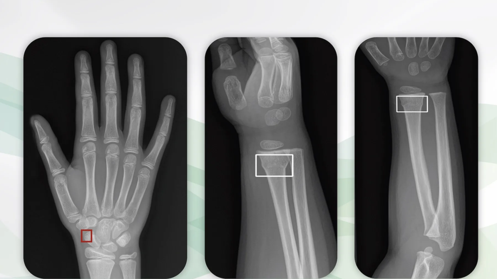
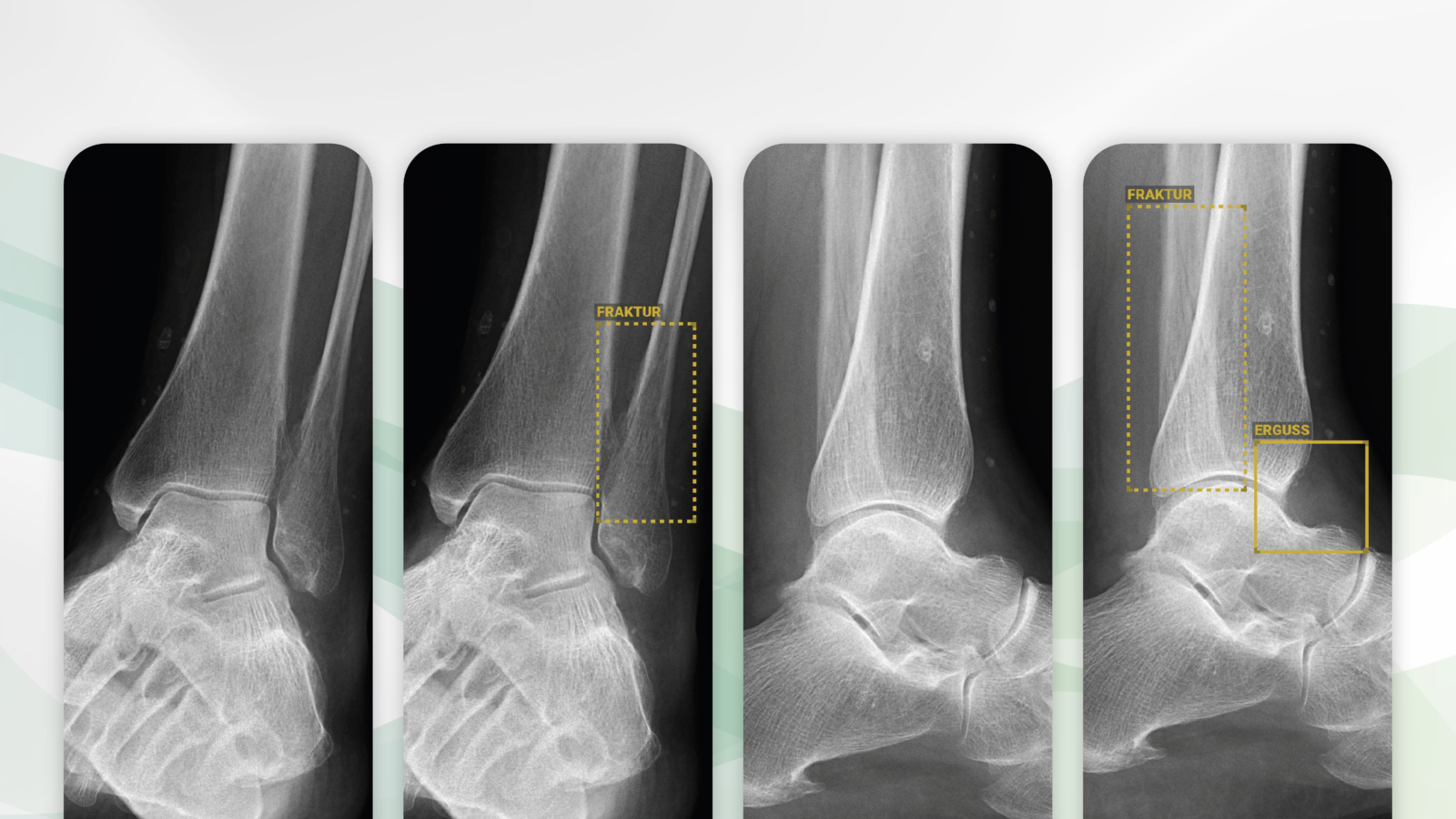
Radiographs of the left ankle frontal (left) and lateral (middle right) views of a 52-year-old patient with pain after a fall. BoneView Trauma correctly detects the nondisplaced longitudinal fracture of the distal fibula (middle left and right, yellow dashed boxes). In addition, the joint effusion is correctly reported in the lateral view (right, yellow box).

Radiographs of the peripheral skeleton are among the most common and important diagnostic methods for medical practices, medical institutes and hospitals. At the same time, they take up a large part of the available working time of radiologists, particularly because on average only about 10% of all radiographs show acute pathologies.

It is with great pleasure that we present BoneView Trauma, an AI assistant for fracture detection on X-ray images.

Why BoneView Trauma matters and how it works

BoneView Trauma detects traumatic lesions on X-rays. The AI diagnoses are available to all patients at any time of the day while providing the convenience and efficiency to streamline workflows. Fractures, effusions, dislocations, dislocations and malignant bone lesions are recognized. These pathologies are detected immediately after the X-rays are taken, even before the radiologist has seen the studies himself. These patients can be prioritized, diagnosed and treated accordingly.

Who benefits

Patients, clinicians and radiologists with a more detailed and accurate diagnosis and the reduced likelihood of missed therapy.

Our own experience at Radailogy

The high true positive rate of BoneView Trauma convinces us. Doubtful or borderline pathologies are also reported as such. In particular, the detection of joint effusions is currently unique. The clear delineation of pathologies in words and images supports the transfer of radiological knowledge to referring physicians and patients.

Any medical professional can request fracture analysis from this AI assistant for any individual patient by uploading it to Radailogy quickly and easily. Our telemedicine customers also use BoneView Trauma as a standard in their daily practice to streamline their workflow.

The scientific evidence

Cohen M, Puntonet J, Sanchez J, Kierszbaum E, Crema M, Soyer P, Dion E. Artificial intelligence vs. radiologist: accuracy of wrist fracture detection on radiographs. Eur Radiol. 2023 Jun;33(6):3974-3983

Duron L, Ducarouge A, Gillibert A, Lainé J, Allouche C, Cherel N, Zhang Z, Nitche N, Lacave E, Pourchot A, Felter A, Lassalle L, Regnard NE, Feydy A. Assessment of an AI Aid in Detection of Adult Appendicular Skeletal Fractures by Emergency Physicians and Radiologists: A Multicenter Cross-sectional Diagnostic Study. Radiology. 2021 Jul;300(1):120-129

Regnard NE, Lanseur B, Ventre J, Ducarouge A, Clovis L, Lassalle L, Lacave E, Grandjean A, Lambert A, Dallaudière B, Feydy A. Assessment of performances of a deep learning algorithm for the detection of limbs and pelvic fractures, dislocations, focal bone lesions, and elbow effusions on trauma X-rays. Eur J Radiol. 2022 Sep;154;110447

Data to upload to Radailogy

Digital radiography of the peripheral skeleton in two planes, for example a.p. and lateral or axial

Radailogy ChestView

Rapid AI diagnosis of the chest X-ray

Chest radiograph of a 39-year-old patient (left) with symptoms of a lower respiratory tract infection. Small lung opacities are visible in projection to the left lower lung lobe. ChestView correctly identifies the pathology (yellow box). The result is also listed in a table (right). In addition to the fully automatic notification of the acute pathology, other threatening pathologies of the thorax are correctly evaluated as negative.

Chest radiography is used worldwide as one of the most common, if not the most common, radiological examinations in both acute care and elective medicine. Correct and reproducible reports and the communication of results from radiologists to patients and clinicians are of the utmost importance for everyone involved.

It is with great pleasure that we present ChestView, an AI assistant for chest radiography.

Why ChestView matters and how it works

ChestView is an expert-level AI assistant that has been extensively tested thanks to the joint work of our multidisciplinary team of developers and radiologist.

With ChestView essential pathologies of the thorax are diagnosed. The AI assistant was developed on the one hand to support triage in acute medicine and on the other hand to improve radiological work in terms of time savings and increased accuracy.

Who benefits

Patients, clinicians and radiologists by identifying the most important chest diseases with pictures and tables.

Our own experience at Radailogy

ChestView supports the detection of major thoracic pathologies. Although pneumonia is referred to as alveolar syndrome, the differential diagnosis has worked with a high degree of accuracy in our detailed tests. The presentation of the results by means of boxes and tabular description is helpful for radiological knowledge transfer to clinicians and patients.

The scientific evidence

Bennani S, Regnard NE, Lassalle L, Nguyen T, Malandrin C, Koulakian H, Khafagy P, Chassagnon G, Revel MP. Evaluation of radiologists’ performance compared to a deep learning algorithm for the detection of thoracic abnormalities on chest X-ray. In press

Data to upload to Radailogy

Digital radiographs of the chest for patients aged 15 and older

Radailogy | AiraMed

Early detection of neurodegenerative diseases for a successful treatment

3D T1-weighted gradient-echo MRI of a 68 years old patient with mild symptoms of cognitive impairment. Routine MRI (not shown) revealed mild microangiopathic neurodegeneration. Brain volumetry was performed (left). AIRAscore identified a volume reduction of the right temporal lobe and bilateral hippocampal that exceeds age (right), suggestive for Alzheimer´s disease. Diagnosis was confirmed by cerebrospinal fluid analysis showing pathological beta amyloid ratio and elevated tau and phosphor-tau proteins.

For many people it remains unrecognized that even minor symptoms can indicate the development of dementia. They thus miss the important early diagnosis and the timely start of their individualized therapy and thus the best possible prognosis for many years. AI, together with brain MRI, can contribute to the earliest possible detection and treatment of neurodegenerative diseases.

It is with great pleasure that we present AIRAscore, an AI assistant for the precise diagnosis of neurodegenerative diseases.

What AIRAscore is and how it works

AIRAscore measures relevant biomarkers for the diagnosis and differential diagnosis of neurodegenerative diseases from MR images of the brain.

Global and regional brain volumes are accurately measured. The results are always compared with age- and gender-specific reference values. The measurements are many times more accurate than even specialists have been able to carry out in the past. Results that deviate from normal are clearly evident. The findings are reported in tabular form in bar graphs and support the knowledge transfer from radiologists to patients and physicians.

Who benefits

Patients, clinicians and radiologists through the early detection and differential diagnosis of neurodegenerative diseases. AIRAscore is also ideal for monitoring the progress of diagnoses made.

Our own experience at Radailogy

We have tested AIRAscore intensively and worked out many technological criteria with the manufacturer for optimal use with Radailogy.

We found a high correlation in the detection of macro- and microangiopathic changes and brain atrophy between our specialists and AIRAscore. In addition, AIRAscore was superior to the human observer in the precision of subtle regional findings in almost all tests. The AI assistant provides accurate volumes of all relevant anatomical structures of the cerebrum, cerebellum and brainstem. Early diagnosis was possible, especially for Alzheimer’s patients, and the correlation with clinical and laboratory data was also very high in the follow-up. AIRAscore can also be used for the differential diagnosis of multiple sclerosis and Parkinson’s disease.

The scientific environment

AIRAscore was developed in neuroscientific medical research. From our point of view, the AI assistant represents a bridge between innovative university research and direct applicability in everyday clinical practice.

Data to upload to Radailogy

1.5-3.0 Tesla MRI, native 3D T1-weighted gradient-echo sequences, slice thickness 1 mm, echo time ≤ 5 ms, flip angle ≤ 15°

Radailogy

Computed tomography: The most important radiological method with improved image quality and reduced radiation dose

Low-dose chest CT of a patient with a pneumonic infiltrate of the middle lobe. Original image with 80kVp, 15 mAs, radiation dose 0.2 mGy (bottom left). Processing with Pixelshine (top left). A comparable image would be created with about 120 kVp, 150 mAs and a radiation dose of about 8 mGy. The radiation dose is reduced by more than 95% with PixelShine.

Low-dose CT of the abdomen. Original image with 120kVp, slice thickness 1.25 mm, radiation dose 1.7 mGy (bottom middle). Processing with Pixelshine (top middle). A comparable image would be created with a radiation dose of about 10 mGy. The radiation dose is reduced by more than 80% with PixelShine.

CT of the brain. Original image with 120kVp, slice thickness 0.625 mm, radiation dose 11 mGy (bottom right). Processing with Pixelshine (top right). A comparable image would be created with a radiation dose of about 40 mGy. The radiation dose is reduced by around 75% with PixelShine.

For several years, medicine has been generating significantly more radiation doses than the natural radiation from the cosmos and the earth ever did. The main reason for this is the constantly increasing radiological use of computed tomography (CT). Precisely because CT is and will remain essential for adequate patient care in almost all diagnostic areas, it is up to us to keep the long-documented radiation-induced cancer risk at the lowest possible level.

It is with great pleasure that we present PixelShine, an AI assistant for CT radiation dose reduction.

Why PixelShine matters

Both in the hospital and in the radiological institute, care is taken to ensure that each patient is only given the necessary radiation dose. However, these low-dose CT protocols almost always produce noisy images, and the CT studies are often difficult to interpret even for medical specialists. In addition, radiologists often have to read CT studies from CT machines from different vendors, which contributes to inconvenience and delays in the workflow.

PixelShine allows two things: Firstly, low-dose CT studies can be carried out for all patients in terms of optimal radiation protection, and PixelShine subsequently generates significantly improved quality from these noisy images, for example in obese patients. Secondly, the lifespan of CT scanners is extended by reducing the load on the CT tubes.

When and how to use PixelShine

PixelShine can be used for studies of any CT device age and vendor. This AI ​​assistant improves radiological precision by homogenizing the workflow.

PixelShine enables radiologists to read noisy CT studies with a high noise level of image noise in the best possible way, and the radiological quality meets the requirements for diagnostic validity.

Furthermore, hospitals and radiological institutes can carry out low-dose CT studies as standard, integrate PixelShine in post-processing and thus achieve consistently high image quality.

Who benefits

Patients, clinicians, radiologists and the management of hospitals and radiological institutes: care for all patients by minimizing the radiation dose, clear CT images, optimal assessment and discussion of findings, money savings by extending the lifespan of CT scanners.

Our own experience at Radailogy

Our customers send us CT studies to improve image quality with PixelShine and enable optimal diagnostic results. Both in individual cases through the simple upload to Radailogy, as well as as a standard in daily cooperation with our telemedicine.

Selection of scientific publications

Hata A, Yanagawa M, Yoshida Y, et al. Combination of Deep Learning–Based Denoising and Iterative Reconstruction for Ultra-Low-Dose CT of the Chest: Image Quality and Lung-RADS Evaluation. American Journal of Roentgenology. 2020;215(6):1321-1328.

Steuwe A, Weber M, Bethge OT, et al. Influence of a novel deep-learning based reconstruction software on the objective and subjective image quality in low-dose abdominal computed tomography. BJR. 2021;94(1117):20200677.

Brendlin AS, Plajer D, Chaika M, et al. AI Denoising Significantly Improves Image Quality in Whole-Body Low-Dose Computed Tomography Staging. Diagnostics. 2022;12(1):225.

Hasegawa A, Ishihara T, Thomas MA, Pan T. Noise reduction profile: A new method for evaluation of noise reduction techniques in CT. Medical Physics. 2022;49(1):186-200.

Nagaraj Y, de Jonge G, Andreychenko A, et al. Facilitating standardized COVID-19 suspicion prediction based on computed tomography radiomics in a multi-demographic setting. Eur Radiol. 2022;32(9):6384-6396.

Hasegawa A, Ishihara T, Thomas MA, Pan T. Noise reduction profile: A new method for evaluation of noise reduction techniques in CT. Medical Physics. 2022;49(1):186-200.

Data to upload to Radailogy

CT studies of any CT scanner age and vendor

Radailogy

Faster diagnoses for the chest X-ray in words and pictures

Chest radiography of a 65 years old patient with symptoms of lower respiratory tract infection. Multiple lung opacities and consolidations are visible in projection to the lower lung lobes as well as the middle lobe. Rayscape correctly identifies all consolidations (red) and opacities (green). Rayscape rates the probability for viral disease as intermediate (3 out of 6) and correctly suggests bronchopneumonia as the differential diagnosis. In addition, some small lung lesions are reported (pink) that were initially missed by the radiologists.

Chest radiography is one of the most common radiological examinations of all, and it is also an essential first-line imaging modality in hospitals and doctors´ offices. The precision in reporting and the communication of results from radiologists to patients and clinicians are the most important success factors for any optimal discussion of findings and therapy.

It is with great pleasure that we present Rayscape, an AI assistant for chest radiography.

What Rayscape is and how it works

Rayscape increases accuracy in detecting 17 major thoracic pathologies.

Thoracic findings are reported and visualized in tabular form. For each finding, the true positive rate is presented as a degree of probability versus differential diagnoses, especially for pulmonary nodules, pneumonia, and mediastinal pathologies. The clear presentation of findings is a welcome support for knowledge transfer from radiologists to patients and doctors.

Who benefits

Patients, clinicians and radiologists by identifying the most important chest diseases and injuries with a clear presentation of the findings in words and pictures.

Our own experience at Radailogy

We tested Rayscape for several years and brought it to life with the manufacturer. We reviewed the performance data and compared it to our own observations:

Rayscape supports the detection of pulmonary nodules, pulmonary consolidation, pulmonary edema, pulmonary emphysema, interstitial lung disease, tuberculosis, pneumothorax, pleural effusion, atelectasis, cardiomegaly, hilar and mediastinal pathologies, diaphragmatic abnormalities, fractures, scoliosis, catheters and drains. From our point of view, Rayscape achieves the best results in the detection of pneumonia, including viral pneumonia with probability grading from 1 to 6, pulmonary nodules, pneumothorax, pleural effusions, cardiac decompensation, consolidations and atelectasis.

The performance

Rayscape has a high level of accuracy in detecting 17 major chest pathologies. Area Under the Receiver Operating Characteristics (AUROC) is highest for tuberculosis (99.1), pneumothorax (97.4), pulmonary edema (94.7), consolidations (94.6), lowest for interstitial lung disease (81, 5), scoliosis (82), diaphragmatic anomalies (85.4), and hilar and mediastinal pathologies (87.7).

Data to upload to Radailogy

Digital radiography of the chest p.a. or a.p.


Radailogy

AI brings patients and doctors together

Physicians depend on exam data from a variety of medical specialties to inform patients about their current situation and enable the best to heal them. 

Radiology and its fast and accurate diagnostics often present itself as the basis on which every successful therapy relies upon. The most important success factors are the precision in the reporting and the transmission of the results to patients and clinicians.

Patients and clinicians rightly want precise analyses presented in words and pictures. X-rays, CT and MR images are ideal for discussing findings. Here, clinicians depend on the results being clearly visible in order to show them to their patients without loss of information or quality.

There has been a 78% increase in radiological examinations since 2008, and the trend is rising. At the same time, healthcare systems are facing a significant shortage of medical specialists. That essential, precise transfer of knowledge from radiologists to patients and clinicians is often subject to daily time pressure.

Radailogy’s AI creates a better future. AI manufacturers know what requirements and expectations we have of their products. We test and perfect AI assistants hand in hand with its developers. At Radailogy, we care about everyone involved: patients, clinicians and radiologists. With every AI assistant we offer, we help save time, increase medical precision, and see and understand exam results clearly.

Radailogy

Optimal lumbar spine MRI visualisation

T2 axial (upper left) and T2 sagittal (lower left) MRI of the lumbar spine of a patient with back pain, lower limb weakness and paresthesia. Left foraminal disc extrusion (red) at the level L4/L5 with impingement of the left L4 nerve (pink) and discrete recessal displacement of the left L5 nerve root (pink). T2 axial (upper right) and T2 sagittal (lower right) MRI of a patient with left lower limb weakness and severe back pain. Left paramedian disc herniation (red), dural sac compression (light blue), recessal displacement of the left L5 nerve root (pink). No foraminal nerve compression (pink). Grade I retrolisthesis.

The MRI of the lumbar spine is one of the most common radiological examinations. Four out of five people will have it at some point in their lives. The most important success factors for any optimal therapy are the precision in reporting and the transmission of the results from radiologists to patients and clinicians.

It is with great pleasure that we present CoLumbo, an AI assistant for MRI of the lumbar spine.

What CoLumbo is and how it works

CoLumbo saves time and increases accuracy in the detection of the most common pathologies of the lumbar spine.

Lumbar spine MRI findings are reported and visualized. It assists in the knowledge transfer from radiologists to both patients and clinicians. In addition, all findings are written in comprehensive, standardized reports and can be used as an integral, automatically filling part of final documents.

Who benefits

Patients, clinicians, and radiologists by more detailed and more accurate diagnosis with subsequent decreased likelihood of suboptimal therapy or surgery. Accurate automatic measurements and clear colorful depiction reduce the need for measuring MRI findings by hand.

Our own experience at Radailogy

We have scrunitely studied CoLumbo over many months. Here we share some of our results with you:

This AI assistant supports the detection of disc herniation, disc bulging, central spinal stenosis, nerve root impingement, reduced vertebral and disk height, hypo- and hyperlordosis as well as spondylolisthesis and pseudolisthesis. With the actual CoLumbo version, we recommend the specialist´s review of foraminal nerve root impingement.

The scientific evidence

CoLumbo´s performance has been tested in clinical research with excellent results in accuracy for intervertebral disc detection and labeling (100%), for the detection of disc herniation (87%; 95% CI: 0.84, 0.89), extrusion (86%; 95% CI: 0.84, 0.89), bulging (76%; 95% CI: 0.73, 0.78), spinal canal stenosis (98%; 95% CI: 0.97, 0.99), nerve root compression (91%; 95% CI: 0.89, 0.92), and spondylolisthesis (87.61%; 95% CI: 85.26, 89.21), respectively.

Lehnen NC, Haase R, JFaber J, Rüber T, Vatter H, Radbruch A, Schmee FC. Detection of Degenerative Changes on MR Images of the Lumbar Spine with a Convolutional Neural Network: A Feasibility Study. Diagnostics 2021; 19;11(5):902

Data to upload to Radailogy

1.0-3.0 Tesla MRI, T2 axial and sagittal 2D and 3D, slice thickness 3.45-5 mm