gleamer upgrade 1 060524

New features for automated measurements of body axes on X-ray images: Part 2

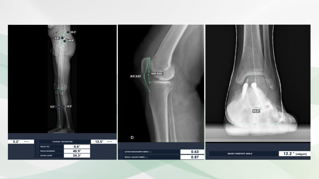
Lateral radiographs of both legs (left) and the right knee joint (middle), radiography of the left ankle and forefoot a.p. (right). BoneView Measurements calculates the iliosacral body axes, the pelvic tilt and the femorotibial joint angles to determine the genu flexum and recurvatum (left). The Caton-Deschamps and Insall-Salvati indices (middle) as well as the Méary hindfoot angle (right) are precisely indicated.

We have the latest upgrade available for you! Gleamer´s BoneView Measurements of body axes come with interesting, new functions.  

What is new?

Caton-Deschamps and Insall-Salvati indices

The Caton-Deschamps index is used to measure patellar height and identify patella alta and patella baja. It relies upon the length of the patellar articular surface and its distance from the tibia.

The Insall-Salvati index is the ratio of the patella tendon length to the length of the patella and is used to determine patellar height.

Genu flexum and Genu recurvatum

Genu flexum describes the pathological reduction of full knee extension, also known as flexion contracture. Normal active range of motion is 0° extension and 140° flexion.

In Genu recurvatum, or back knee, the knee bends backwards. In this deformity, excessive extension of the femorotibial joint occurs.

Hindfoot angle on Méary´s views

Méary view X-rays measure the angular deviation of the hindfoot axis in order to assess hindfoot malalignment.

The scientific evidence

van Duijvenbode D, Stavenuiter M, Burger B, van Dijke C, Spermon J, Hoozemans M. The reliability of four widely used patellar height ratios. Int Orthop. 2016 Mar;40(3):493–497.

Kadakia N & Ilahi O. Interobserver Variability of the Insall-Salvati Ratio. Orthopedics. 2003;26(3):321-323.

Neri, R. Barthelemy, Y. Tourné. Radiologic analysis of hindfoot alignment: Comparison of Méary, long axial, and hindfoot alignment views. Revue de Chirurgie Orthopédique et Traumatologique. 2017;103(8):882-887.

Data to upload to Radailogy

Digital radiography of the skeleton

gleamer upgrade 2 060524

New features for automated measurements of body axes on X-ray images: Part 1

Radiography of the pelvis and both hip joints of a boy with the acetabular angles precisely measured on both sides by the AI ​​assistant (left).

Frontal radiography of both forefeet of a 55-year-old patient. On the right (on the left in the picture) there are normal joint angles of the forefoot after osteomy of the first metatarsal bone. There is a hallux varus deviation on the left (on the right in the picture). The AI ​​assistant measures the respective joint angles accurately.

We have the latest upgrade available for you! Gleamer´s BoneView Measurements of body axes come with interesting, new functions.

What is new?

Risser index

The Risser index is used to grade skeletal maturity for patients up to 18 years of age based on the degree of ossification and fusion of the iliac crest apophyses. It is primarily used as a marker of skeletal maturity, a surrogate for growth rate and potential, to plan scoliosis correction.

Acetabular angle for congenital hip measurement

The acetabular angle, or Sharp’s angle, is used to assess developmental hip dysplasia, particularly in patients with already ossifying epiphyses and therefore limited sonographic assessment.

Lumbar spine

The performance on lateral view measurements of the lumbar spine has significantly improved. Furthermore, lordosis is now calculated between L1 and S1, instead of between L1 and L5.

The scientific evidence

Hacquebord J, Leopold S. In Brief: The Risser Classification: A Classic Tool for the Clinician Treating Adolescent Idiopathic Scoliosis. Clin Orthop Relat Res. 2012;470(8):2335-2338.

Lee Y, Chung C, Koo K, Lee K, Kwon D, Park M. Measuring Acetabular Dysplasia in Plain Radiographs. Arch Orthop Trauma Surg. 2011;131(9):1219-1226.

Data to upload to Radailogy

Digital radiography of the skeleton