
Radailogy
Simply Intelligent
Simply Intelligent
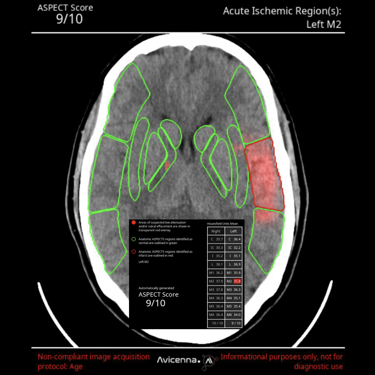
Brainscan CT reports acute and chronic intracranial processes in CT scans using tables and heat maps.

CINA-IPE detects acute pulmonary artery embolism in CT angiographies.
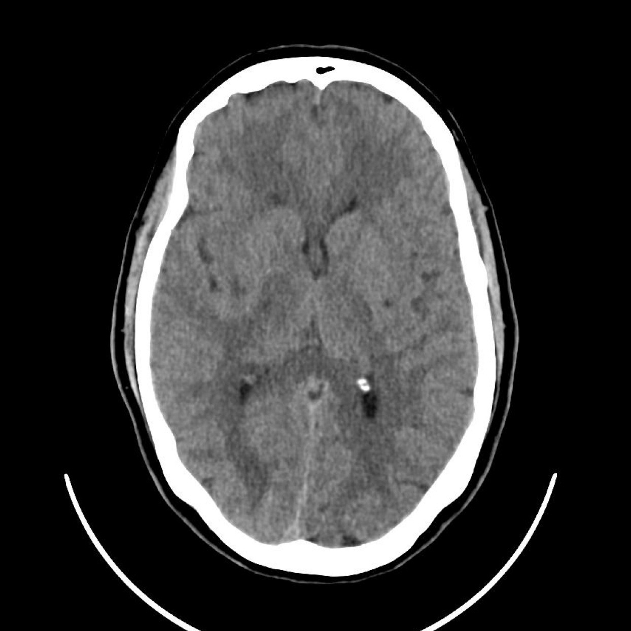
CINA-ICH detects acute intracranial hemorrhage in cerebral CT studies.
CINA-LVO detects acute intracranial artery occlusion in CT angiographies.
PixelShine generates significantly improved quality from these noisy images, for example in obese patients. Secondly, the lifespan of CT scanners is extended by reducing the load on the CT tubes.