
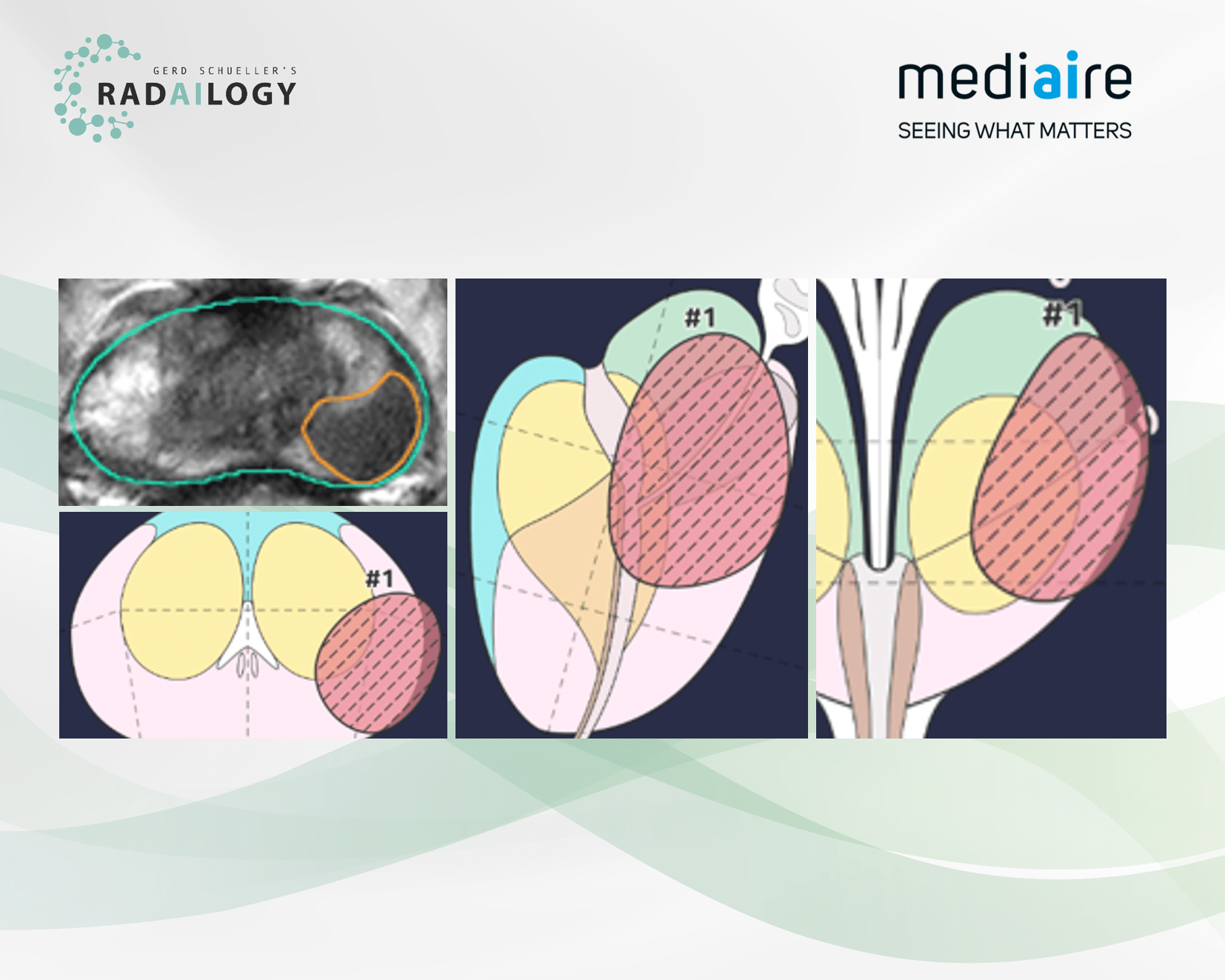
MRI of the prostate of a 59-year-old patient with a Gleason score of 4. T2 hypointensity in the peripheral zone on the left with corresponding hyperintense DWI signal and elevated ADC values (not shown). The lesion is detected by mdprostate, scored as PI-RADS 5, and displayed in 3D images (axial, lower left; coronal, middle; sagittal, right). The volume is calculated to be 2.7ml, the dimensions are 17 x 13.3 x 24mm. Histopathology confirmed the lesion malignant.
Men often hear that prostate cancer is very common. Only radiologists with extensive experience in oncology are able to distinguish between benign and malignant lesions. And if the findings are inconclusive, is there any useful support available? Yes, there is!
We are pleased to present mdprostate, an AI assistant for the detection and analysis of prostate tumors in MR studies.
Why mdprostate matters and how it works
In brief: prostate tumors are localized, measured, and evaluated using the PI-RADS 2.1 classification system. The results are presented in three-dimensional color graphics and clear tables. The automated comparison with preliminary studies is particularly interesting.
Who benefits
Any man seeking a second opinion. And, of course, clinicians and radiologists benefit from the detailed presentation of prostate tumors with clear images and tables.
Our own experience at Radailogy
Our tests revealed a high TP rate of >80% and a TN rate of >85%. The sensitivity in our sample was 87%, and the specificity was 69%. The score is based on the PI-RADS 2.1 classification. mdprostate also calculates prostate volume. We were impressed by the detailed depiction of the prostate with comprehensive tables and 3D images.
Any man can request an AI-assisted second opinion for his prostate MRI by quickly and easily uploading prostate MRI studies to Radailogy. Our telemedicine clients also use this AI assistant as a standard in their daily practice to optimize their oncology workflow.
The scientific evidence
Bayerl N, Adams LC, Cavallaro A, Bäuerle T, Schlicht M, Wullich B, Hartmann A, Uder M, Ellmann S. Assessment of a fully-automated diagnostic AI software in prostate MRI: Clinical evaluation and histopathological correlation. Eur J Radiol. 2024 Dec;181:111790
Data to upload to Radailogy
1.5-3.0 Tesla; T2w SE: FOV 10-12mm; imaging resolution 0.7-0.9mm; slice thickness 3-4mm; interval <0.5mm; DWI: b value 1400-2000; FOV 14-16mm; imaging resolution 2.1-2.5mm; slice thickness 3-4mm; interval <0.5mm; ADC: ; FOV 14-16mm; imaging resolution 2.1-2.5mm; slice thickness 3-4mm; interval <0.5mm