
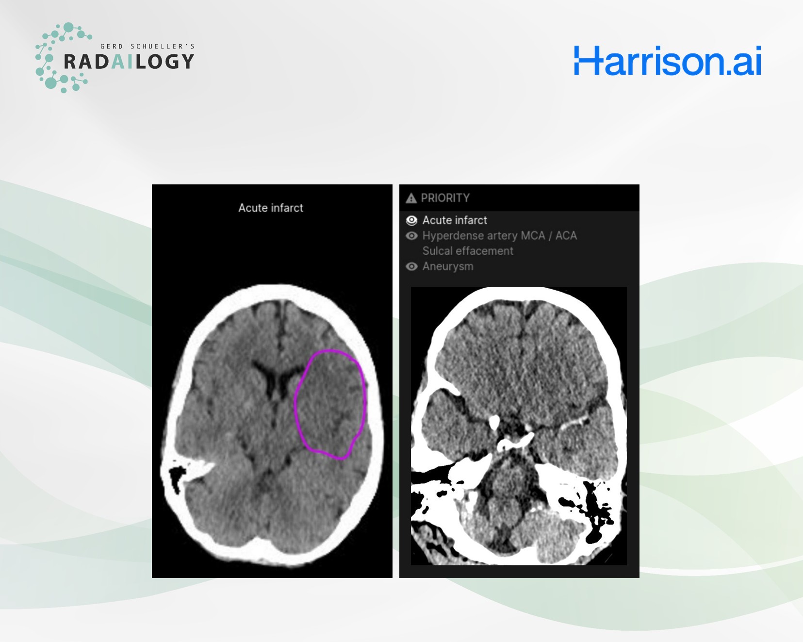
Non-enhanced cranial CT of a 61-year-old patient with acute right-sided hemisymptoms. Harrison Brain CT detects acute left parietal ischemia (left) and acute thrombosis of the middle cerebral artery (right). The pathologies are listed in a table and displayed directly in the CT images.
We need reliable AI assistants in neurology and traumatology. A new tool for native cranial CT helps detect acute and chronic lesions.
It is with great pleasure that we present Harrison CT Brain, an AI assistant for cranial CT studies.
How Harrison Brain CT works
The AI assistant reports acute and chronic intracranial processes in native CT scans. The lesions are tabulated with emphasis on their acuity. Most pathologies, at least the acute ones, are drawn directly into the CT images as colored annotations.
Any medical professional can use Harrison CT Brain by quickly and easily uploading cranial CT studies to Radailogy.
Who benefits
Patients, clinicians and radiologists through the detailed listing of cranial pathologies with tables and the direct annotation in CT images.
Our own experience at Radailogy
In our first series of tests, we saw precise results, particularly in acute intracranial hemorrhages and fractures of the facial and cerebral skull. We found the distinction between acute and chronic parenchymal lesions encouraging. Our data on sensitivity and specificity of approximately 80% for acute bleeding were comparable to those of the available publications. The manufacturer’s list of 130 detectable pathologies needs to be verified in larger studies. The decision to use the AI assistant as a triage tool in teleradiology also requires comprehensive data. We are currently conducting such a prospective study. We’ll keep you updated.
The scientific evidence
Hillis JM, Bizzo BC, Newbury‐Chaet I, Mercaldo SF, Chin JK, Ghatak A, Halle MA, L’Italien E, et al. Evaluation of an Artificial Intelligence Model for Identification of Intracranial Hemorrhage Subtypes on Computed Tomography of the Head. Stroke Vasc Interv Neurol. 2024 May 16;4(4):e001223
Data to upload to Radailogy
Non-enhanced cranial CT studies of any CT scanner for patients aged 18 and older; axial reformations; maximal slice thickness 1.5 mm; soft tissue reconstruction kernel