
Radailogy
Simply Intelligent
Simply Intelligent
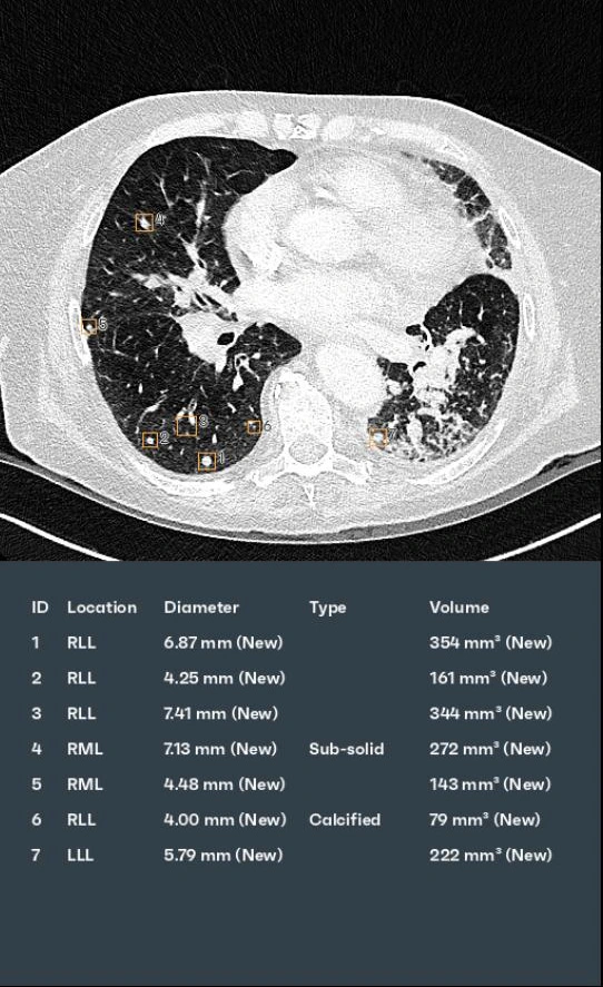
aview LCS detects and diagnoses lung nodules on chest CT studies. An important function is the automated comparison of nodules in the CT follow up.
aview COPD classifies and analyses COPD patterns on chest CT studies. An important feature is the tracking of disease in follow up CT studies.
PixelShine generates significantly improved quality from these noisy images, for example in obese patients. Secondly, the lifespan of CT scanners is extended by reducing the load on the CT tubes.
aview CAC analyzes calcium in the coronary arteries and calculates the risk of a heart attack.