Prostate Intelligence helps detect and analyze prostate cancer in MR studies. The rating of the results is based on the PI-RADS classification. The findings are clearly structured, and enable the selection of biopsy targets with high-quality segmentations.
Prostate Intelligence presents the results in a structured manner with heat maps and tables. These features are a welcome aid for radiologists in the detection and evaluation of prostate tumors. In addition, the AI assistant saves radiologists and clinicians considerable time in organ and tumor segmentation, and the interdisciplinary case discussion increases in quality.
1.5-3.0 Tesla, imaging planes should be the same across T2, DWI and DCE; T2: recommended slice thickness 3mm, imaging resolution <0.65mm; DWI & ADC: TE ≤90 msec; TR ≥3000 msec, slice thickness ≤4mm, no gap, resolution ≤2.5mm phase and frequency, high b-value ideally 800-1000; Contrast enhanced T1 (DCE): TR/TE <100msec 2min. Ideally the enhancement peak should occur at least 1min before observations stop. There is no need for DCE observations longer than 3min.
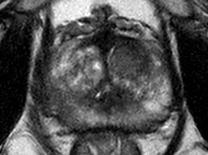
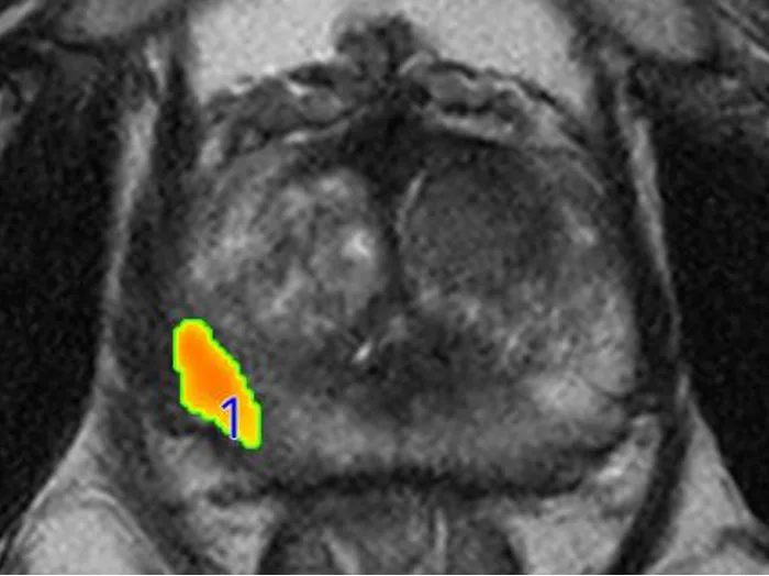
Axial T2 MRI of the prostate of a 63-year-old patient with a Gleason Score 4. T2 hypointensity in the right middle peripheral zone (left) with corresponding hyperintense DWI signal and marked hypervascularization (not shown). Prostate Intelligence has detected (overlaid to T2 axial) and rated the lesion as AI-Likert 4. Prostate Intelligence quantified the prostate volume as 68 ml (not shown). Histopathology confirmed the lesion malignant.
Similar AI Assistants
mdprostate
mdprostate detects and analyzes prostate tumors in MR studies