The diagnosis of fractures, effusions, dislocations and malignant bone lesions is supported. The clear presentation of the results in words and images supports the radiological knowledge transfer to referring physicians and patients.
With BoneView Trauma, both time savings and increased diagnostic certainty can be achieved. Patients with detected pathologies can be prioritized and treated accordingly.
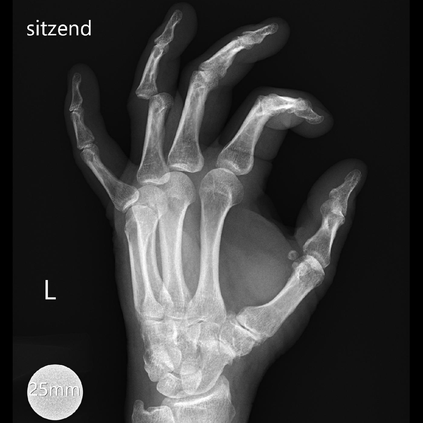
Digital radiography of the peripheral skeleton in two planes, for example a.p. and lateral or axial
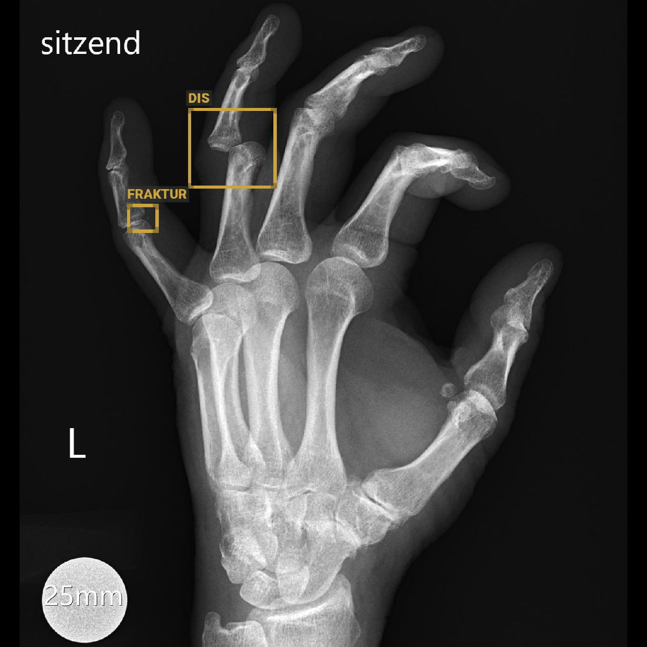
Radiograph of the left hand of a 65-year-old man with pain after a fall. BoneView Trauma correctly detects the ulnar dislocation of the middle phalanx of the proximal interphalangeal joint of the ring finger. In addition, a non-displaced fracture of the base of the middle phalanx of the little finger, which the radiologists did not describe, is correctly reported.
Similar AI Assistants
BoneView Bone Age
BoneView Bone Age calculates bone age using hand radiography.